Galerie d'images
Toutes les images de la base — taxons, formations et intervalles géologiques.
⚠ La fonctionnalité de récupération des images est en cours de test, des images non pertinentes peuvent apparaître.
2,142 image(s)

Cimoliopterus cuvieri. Holotype NHMUK PV 39409 (Cenomanian / Turonian, Chalk Formation), anterior part of the rostrum A right lateral view B respective line drawing C ventral view D respective line drawing. Abbreviations: m – maxillae, pm – premaxillae, pmcr – premaxillary crest, prid – palatal ridge. Arrows and numbers indicate alveoli or teeth and their respective position. Scale bar = 10 mm. Photos courtesy of The Natural History Museum.


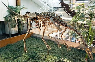
Fossil specimen of early Cretaceous pterosaur Sinopterus dongi, which is collected from Chaoyang, Liaoning, China. The specimen (BMNHC Ph773) is a collection of Beijing Museum of Natural History and was on display in the National Museum of Natural Science (Taichung, Taiwan) during a special exhibition.
Fossil specimen of early Cretaceous pterosaur Sinopterus dongi, which is collected from Chaoyang, Liaoning, China. The specimen (BMNHC Ph773) is a collection of Beijing Museum of Natural History and was on display in the National Museum of Natural Science (Taichung, Taiwan) during a special exhibition.


Camposipterus nasutus comb. n. Holotype CAMSM B 54556 (Albian, Cambridge Greensand), anterior part of the rostrum A left lateral view B respective line drawing C ventral view D respective line drawing. Abbreviations: m – maxillae, pm – premaxillae, prid – palatal ridge. Arrows and numbers indicate alveoli or teeth and their respective position. Scale bar = 10 mm.

Holotype skeleton of Jianchangnathus robustus on display at the Paleozoological Museum of China.


Skeletal reconstruction of Limusaurus inextricabilis. Matches proportions in published skeletal reconstruction by Gregory S. Paul in the 2016 book The Princeton Field Guide to Dinosaurs.


Holotype of Ulughbegsaurus as well as the holotype placed on a reconstruction of Ulughbegsaurus's skull


A diagram of the dromaeosaurid dinosaur Pyroraptor olympius with selected fossil elements that can be reliably scaled using measurements given by Allain & Taquet (2000) to produce a realistic estimate of the taxon's size and proportions in life. This silhouette is based on the supposition of the missing elements of Pyroraptor's skeleton having fairly generalized dromaeosaurid proportions.

Buitreraptor displayed at the Royal Ontario Museum, Toronto, Canada.

Sinornithosaurus millenii fossil displayed in Hong Kong Science Museum

Cast of the known material of Acheroraptor, on display at the Smithsonian's National Museum of Natural History's exhibit The Last American Dinosaurs.

Fossiles de Variraptor présentés lors de l'exposition "Sur les traces des dinosaures de la montagne Sainte-Victoire"

Fossil specimen of Anchiornis huxleyi on display at the Beijing Museum of Natural History.

Skeletal restoration of Byronosaurus jaffei. Scale bar = 10cm

Title: The dinosaur book : the ruling reptiles and their relatives Identifier: bookruli00colb (find matches) Year: 1951 (1950s) Authors: Colbert, Edwin H. (Edwin Harris), 1905-2001; Knight, Charles Robert, 1874-1953; American Museum of Natural History Subjects: Dinosaurs; Reptiles, Fossil Publisher: New York : Published for the American Museum of Natural History by McGraw-Hill Contributing Library: American Museum of Natural History Library Digitizing Sponsor: IMLS / LSTA / METRO View Book Page: Book Viewer About This Book: Catalog Entry View All Images: All Images From Book Click here to view book online to see this illustration in context in a browseable online version of this book. Text Appearing Before Image: ' Text Appearing After Image: bone A typical example of the parts usually found fossilized: portions of the skeleton of a small dinosaur from Mongolia A.M.N.H. photographs One of the rarest fossils: a dino- saur egg over 60 million years old, compared with a hen's egg (left) and an alligator egg (right) Note About Images Please note that these images are extracted from scanned page images that may have been digitally enhanced for readability - coloration and appearance of these illustrations may not perfectly resemble the original work.
Title: The dinosaur book : the ruling reptiles and their relatives Identifier: bookruli00colb (find matches) Year: 1951 (1950s) Authors: Colbert, Edwin H. (Edwin Harris), 1905-2001; Knight, Charles Robert, 1874-1953; American Museum of Natural History Subjects: Dinosaurs; Reptiles, Fossil Publisher: New York : Published for the American Museum of Natural History by McGraw-Hill Contributing Library: American Museum of Natural History Library Digitizing Sponsor: IMLS / LSTA / METRO View Book Page: Book Viewer About This Book: Catalog Entry View All Images: All Images From Book Click here to view book online to see this illustration in context in a browseable online version of this book. Text Appearing Before Image: ' Text Appearing After Image: bone A typical example of the parts usually found fossilized: portions of the skeleton of a small dinosaur from Mongolia A.M.N.H. photographs One of the rarest fossils: a dino- saur egg over 60 million years old, compared with a hen's egg (left) and an alligator egg (right) Note About Images Please note that these images are extracted from scanned page images that may have been digitally enhanced for readability - coloration and appearance of these illustrations may not perfectly resemble the original work.

Skeleton identified as Nemegtomaia by Greg Funston.[1] Central Museum of Mongolian Dinosaurs, Ulaanbaatar. Complete indexed photo collection at WorldHistoryPics.com.

Skeletal reconstruction of alvarezsaurid MPCN-PV 738, referred to Bonapartenykus ultimus.


Teratophoneus curriei, adult (left) and juvenile (right), on display at the Natural History Museum of Utah, Salt Lake City.


Tyrannosauripus pillmorei, probable Tyrannosaurus footprint from w:Philmont Scout Ranch, New Mexico
Tyrannosauripus pillmorei, probable Tyrannosaurus footprint from w:Philmont Scout Ranch, New Mexico

3D model of first track of trackway shown. Left. Orthofoto; right: Heightmap
Mounted specimen of Archaeornithomimus asiaticus on display at the Paleozoological Museum of China.


Reconstructed skull of Einiosaurus procurvicornis on display at the Natural History Museum of Los Angeles County.


Montanoceratops cerorhynchus (Brown & Schlaikjer, 1942) - fossil ceratopsian dinosaur skeleton from the Cretaceous of Montana, USA. (MOR 542, Museum of the Rockies, Bozeman, Montana, USA) The species name is sometimes incorrectly spelled "cerorhynchos". The original publication spells it "cerorhynchus". The genus name is sometimes incorrectly spelled "Montanaceratops". Ceratopsians are the "horned dinosaurs". They were large, quadrupedal, herbivorous dinosaurs having a beaked skull and a frill - an extension of bone behind the skull that partially covered the neck. Ceratopsian dinosaurs are known from the Jurassic and Cretaceous. The last members of the group died out at the Cretaceous-Tertiary boundary, 65 million years ago. This is a partial skeleton of a juvenile Montanoceratops, a ceratopsian from the near-latest Cretaceous of western North America. This type of ceratopsian lacked facial horns. From exhibit signage: Sixty-eight million years ago, when the horned dinosaurs Triceratops and Torosaurus inhabited the coastal plain near the inland ocean, primitive "horned" dinosaurs named Montanoceratops lived in uplands near the young Rocky Mountains. These little protoceratopsians fed on plants with slicing teeth and narrow beaks similar to their giant three-horned relatives. Classification: Animalia, Chordata, Vertebrata, Reptilia, Archosauria, Dinosauria, Ornithischia, Marginocephalia, Ceratopsia, Leptoceratopsidae Stratigraphy: St. Mary River Formation, Maastrichtian Stage, Upper Cretaceous Locality: Little Rocky Coulee, north of the town of Cut Bank, eastern Glacier County, northwestern Montana, USA Info. at: en.wikipedia.org/wiki/Montanoceratops


Skeletal mount of Archaeoceratops, housed in Tamba Dinosaur Museum
Skeletal mount of Archaeoceratops, housed in Tamba Dinosaur Museum


Specimens of Galleonosaurus dorisae n. gen. n. sp. from the Flat Rocks Sandstone in the upper Barremian, Wonthaggi Formation, Gippsland Basin, southeastern Australia: (1–2) holotype (NMV P229196), left maxilla in lateral (1) and medial (2) views; (3) NMV P208178, left maxilla in lateral view; (4) NMV P212845, left maxilla in lateral view; (5) NMV P209977, left maxilla in lateral view; (6) NMV P186440, left maxilla in lateral view; (7) NMV 208113, right maxillary tooth in labial view. Scale bars = 10 mm (1–6); 1 mm (7).

Muttaburrasaurus The plants, animals and climate of the Australian continent have changed dramatically over long periods of time. Imagine this giant creature roaming the luxuriant wet forests that covered parts of the continent in the Cretaceous period, about 100-110 million years ago. The Muttaburrasaurus ambled along on all four legs or stood on its hind legs. Its large teeth were well adapted to eat tough vegetation such as the leathery foliage of the evergreen forests of Araucaria trees, ancient relatives of the bunya pine of south-eastern Queensland. In 1963, grazier Doug Langdon discovered the fossilised bones of a dinosaur on his property near Muttaburra in central-west Queensland. It was one of the most complete dinosaur skeletons found in Australia. The bones belonged to a new species of ornithopod and palaeontologists named it Muttaburrasaurus langdoni. Cast of Muttaburresaurus langdoni 1987 made by Queensland Museum, Brisbane National Museum of Australia


Partial skeleton of Claosaurus agilis (holotype YPM 1190). (A) Right ilium in lateral view. (B) Partial postorbital in lateral view. (C) Distal process of the right ischium in lateral view. (D) Mounted partial skeleton of YPM 1190. (E) Coronoid process of the right dentary in lateral view. (F) Fragment of maxilla in lateral view. (G) Detail of the maxillary tooth crowns in (F). (H) Fragment of maxilla in lateral view. (I) Detail of a maxillary tooth crown in (H).


