←

os

Partie anatomique

85 image(s) · 61 Actualités

Galerie d'images

Entire skeleton of Jidapterus edentus (RCPS-030366CY) preserved in a slab mostly in right lateral view.

Abbreviations: aa, the atlas-axis complex; bo, basioccipital; br, braincase; cve, cervical vertebrae; cv3, the third cervical vertebra; cv8, the eighth cervical vertebra; ds, dentaries; dv, dorsal vertebra; dve, dorsal vertebrae; ep, epiphysis; j, jugal; lc, left carpus; lf, left femur; li, left ilium; lis, left ischium; lm, left manual digits; lp, left pubis; lpe, left pes; lpt, left pteroid; lr, left radius; lsc, left scapula-coracoid; lt, left tibia; lta, left tarsus; lu, left ulna; lwmc, left wing metacarpal; lwp1–4; left wing phalanges 1–4; p, pterygoid; pp, postacetabular process of the right ilium; q, quadrate; rc, right carpus; rh, right humerus; rf, right femur; rfi, right fibula; ribs, ribs; ris, right ischium; rm, right manual digits; rmcI, right metacarpal I; rp, right pubis; rpe, right pes; rpt, right pteroid; rr, right radius; rsc, right scapula-coracoid; rt, right tibia; ru, right ulna; rwmc, right wing metacarpal; rwp1–4, right wing phalanges 1–4; rostrum, rostrum; st, sternum; sv, sacral vertebra; sve, sacral vertebrae. Gray shaded areas bound by solid lines indicate preserved bones, dashed lines indicate bones with margins discerned by impression, and lack of lines indicate missing bone or displacement along cracks in the slab.
Taxons Chaoyangopterinae

Entire skeleton of Jidapterus edentus (RCPS-030366CY) preserved in a slab mostly in right lateral view. Abbreviations: aa, the atlas-axis complex; bo, basioccipital; br, braincase; cve, cervical vertebrae; cv3, the third cervical vertebra; cv8, the eighth cervical vertebra; ds, dentaries; dv, dorsal vertebra; dve, dorsal vertebrae; ep, epiphysis; j, jugal; lc, left carpus; lf, left femur; li, left ilium; lis, left ischium; lm, left manual digits; lp, left pubis; lpe, left pes; lpt, left pteroid; lr, left radius; lsc, left scapula-coracoid; lt, left tibia; lta, left tarsus; lu, left ulna; lwmc, left wing metacarpal; lwp1–4; left wing phalanges 1–4; p, pterygoid; pp, postacetabular process of the right ilium; q, quadrate; rc, right carpus; rh, right humerus; rf, right femur; rfi, right fibula; ribs, ribs; ris, right ischium; rm, right manual digits; rmcI, right metacarpal I; rp, right pubis; rpe, right pes; rpt, right pteroid; rr, right radius; rsc, right scapula-coracoid; rt, right tibia; ru, right ulna; rwmc, right wing metacarpal; rwp1–4, right wing phalanges 1–4; rostrum, rostrum; st, sternum; sv, sacral vertebra; sve, sacral vertebrae. Gray shaded areas bound by solid lines indicate preserved bones, dashed lines indicate bones with margins discerned by impression, and lack of lines indicate missing bone or displacement along cracks in the slab.

os humérus vertèbre Chaoyangopteridae +3
Vertebrae, scapula, forelimb bones, and pelvic bones of Martharaptor greenriverensis (UMNH VP 21400). (A)–Partial cervical neural arch, dorsal view. (B–E)–Cranial dorsal centrum in cranial (B), caudal (C), right lateral (D), and left lateral (E) views. (F–G)–Distal caudal centrum in lateral (F) and ventral (G) views. (H)–Possible ulna. (I)–Possible radius. (J–K)–Left scapula in lateral (J) and medial (K) views. (L)–Proximal end of ischium. (M)–Possible distal end of pubis Scale bar = 50 mm. acr  =  acromium process, ar  =  acetabular rim, gl  =  glenoid, hyp  =  hypapophysis, poz  =  postzygapophysis, pnp  =  pneumatopore, prz  =  prezygapophysis.

Vertebrae, scapula, forelimb bones, and pelvic bones of Martharaptor greenriverensis (UMNH VP 21400). (A)–Partial cervical neural arch, dorsal view. (B–E)–Cranial dorsal centrum in cranial (B), caudal (C), right lateral (D), and left lateral (E) views. (F–G)–Distal caudal centrum in lateral (F) and ventral (G) views. (H)–Possible ulna. (I)–Possible radius. (J–K)–Left scapula in lateral (J) and medial (K) views. (L)–Proximal end of ischium. (M)–Possible distal end of pubis Scale bar = 50 mm. acr  =  acromium process, ar  =  acetabular rim, gl  =  glenoid, hyp  =  hypapophysis, poz  =  postzygapophysis, pnp  =  pneumatopore, prz  =  prezygapophysis.

os écaille Martharaptor partiel
Bones referred to Ibirania parva, A. Middle caudal vertebra (MPMA 08-0060-07) referred to I. parva. B. Views of partial right ulna (LPP-PV-0202) of the I. parva holotype. According to Navarro et al. (2022).

Bones referred to Ibirania parva, A. Middle caudal vertebra (MPMA 08-0060-07) referred to I. parva. B. Views of partial right ulna (LPP-PV-0202) of the I. parva holotype. According to Navarro et al. (2022).

os vertèbre holotype Ibirania +1
Herbivorous dinosaur found in the Al-khoudh area.  This dinosaur is similar to the Zalmoxes and Rhabdodon dinosaurs.  The skeleton in the Bait Al Baranda Museum was assembled from bones borrowed from several museums.

Herbivorous dinosaur found in the Al-khoudh area. This dinosaur is similar to the Zalmoxes and Rhabdodon dinosaurs. The skeleton in the Bait Al Baranda Museum was assembled from bones borrowed from several museums.

os musée Dinosauria Mochlodon +3
Dinosaur Bones

Dinosaur Bones

os Sri Lanka Anzu Dinosauria
MRF 319, a partial oviraptorosaurian skeleton referred to Anzu wyliei. 
(A) Skeletal reconstruction in left lateral view, with preserved bones in gray and bones represented in other Anzu specimens in white (hatching indicates heavily reconstructed portions of the ilia of CM 78001). Middle-posterior (ninth?) cervical vertebra in (B) anterior, (C) left lateral, and (D) dorsal views. Posterior (11th?) cervical vertebra in (E) anterior, (F) left lateral, and (G) dorsal views. Posterior (12th?) cervical vertebra in (H) anterior, (I) left lateral, and (J) dorsal views. Anteroposteriorly crushed left radius in lateral (K) and anterior (L) views. Mediolaterally crushed left ulna in lateral (M) and anterior (N) views. (O) Partial left scapulocoracoid in lateral view. Dorsal rib in anterior (P) and posterior (Q) views. Abbreviations: acr, acromial process; cr, cervical rib; dip, distal processes; pat, pathology; pf, pneumatic fossa. Scale bars = 50 cm in A; 1 cm in B–Q.

MRF 319, a partial oviraptorosaurian skeleton referred to Anzu wyliei. (A) Skeletal reconstruction in left lateral view, with preserved bones in gray and bones represented in other Anzu specimens in white (hatching indicates heavily reconstructed portions of the ilia of CM 78001). Middle-posterior (ninth?) cervical vertebra in (B) anterior, (C) left lateral, and (D) dorsal views. Posterior (11th?) cervical vertebra in (E) anterior, (F) left lateral, and (G) dorsal views. Posterior (12th?) cervical vertebra in (H) anterior, (I) left lateral, and (J) dorsal views. Anteroposteriorly crushed left radius in lateral (K) and anterior (L) views. Mediolaterally crushed left ulna in lateral (M) and anterior (N) views. (O) Partial left scapulocoracoid in lateral view. Dorsal rib in anterior (P) and posterior (Q) views. Abbreviations: acr, acromial process; cr, cervical rib; dip, distal processes; pat, pathology; pf, pneumatic fossa. Scale bars = 50 cm in A; 1 cm in B–Q.

os écaille vertèbre pathologie +5
MRF 319, a partial oviraptorosaurian skeleton referred to Anzu wyliei. Skeletal reconstruction in left lateral view, with preserved bones in gray and bones represented in other Anzu specimens in white (hatching indicates heavily reconstructed portions of the ilia. Scale bar = 50 cm (19.7 in)

MRF 319, a partial oviraptorosaurian skeleton referred to Anzu wyliei. Skeletal reconstruction in left lateral view, with preserved bones in gray and bones represented in other Anzu specimens in white (hatching indicates heavily reconstructed portions of the ilia. Scale bar = 50 cm (19.7 in)

os écaille spécimen Anzu +3
Osteohistology of UALVP 59606, metatarsal IV of Citipes elegans. Overview of histological thin section of UALVP 59606 in plane-polarized (A) and cross-polarized (B) light, showing locations of close-up images. Detail (C) of cortex showing predominantly primary fibrolamellar bone with longitudinal-reticular vascularity, endosteal layer, and cyclical growth marks (arrows), under plane-polarized light. Close-up (D) of well developed endosteal layer on the medullary cavity, showing sec- ondary excavation by simple vascular canals, under cross-polarized light. Close-up (E) of tightly packed cyclical growth marks (arrows) near the periosteal surface of the cortex, under plane-polarized light. Close-up (F) of periosteal surface of cortex, showing avascular parallel-fibered bone indicating an incipient external fundamental system (double ended arrow) with three LAGs (arrows). Abbreviations: efs, incipient external fundamental system; el, endosteal lamellae; hb, Haversian bone; pfb, parallel-fibered bone; radv, radial simple vascular canal; so, secondary osteon.

Osteohistology of UALVP 59606, metatarsal IV of Citipes elegans. Overview of histological thin section of UALVP 59606 in plane-polarized (A) and cross-polarized (B) light, showing locations of close-up images. Detail (C) of cortex showing predominantly primary fibrolamellar bone with longitudinal-reticular vascularity, endosteal layer, and cyclical growth marks (arrows), under plane-polarized light. Close-up (D) of well developed endosteal layer on the medullary cavity, showing sec- ondary excavation by simple vascular canals, under cross-polarized light. Close-up (E) of tightly packed cyclical growth marks (arrows) near the periosteal surface of the cortex, under plane-polarized light. Close-up (F) of periosteal surface of cortex, showing avascular parallel-fibered bone indicating an incipient external fundamental system (double ended arrow) with three LAGs (arrows). Abbreviations: efs, incipient external fundamental system; el, endosteal lamellae; hb, Haversian bone; pfb, parallel-fibered bone; radv, radial simple vascular canal; so, secondary osteon.

os croissance Citipes Leptorhynchos +1
Main evolutionary steps proposed for the morphofunctional and postural changes of the sauropod pedes. (A) Sauropod body mass through time (in metric tons) based on the sauropod body mass estimations of (41) (NB: data lacking for the second half of the Upper Cretaceous so illustrated here faded, in continuity with the data recorded in the Cretaceous). Schematic outlines of selected large specimens illustrated in the curve, including (from left to right) P. engelhardti, Vulcanodon karibaensis, R. brownei, G. brancai, Cedarosaurus weiskopfae, and Notocolossus gonzalezparejasi. (B) Projected evolutionary changes occurring in the sauropod pes associated with trend in body mass, including 1, skeletal and functional digitigrade pedal posture among basal non-sauropod sauropodomorphs with an incipient soft tissue pad (ISP) (see figs. S34 and S35); 2 and 3, expansion of a well-developed soft tissue pad beneath the elevated pedal bones (SP), resulting in a functionally plantigrade pes + retention of skeletal posture within a range of digitigrady; 4, retention of a soft tissue pad and yet undetermined trend toward more elevated bones; 5, conservation of the neomorphic soft tissue pad within all lineages. Selected examples of well-preserved non-sauropod sauropodomorph and sauropod pedal tracks illustrated above the trends, including (from left to right) Evazoum siriguii; Pseudotetrasauropus bipedoida, Eosauropus isp., Lavinipes cheminii; Kalosauropus pollex, Liujianpus shunan, Polyonyx gomesi; Parabrontopodus mcintoshi; Brontopodus birdi; Titanopodus mendozensis; and unnamed Asian sauropod track. Source of adapted drawing and notes are listed in table S9 and data S2.
Taxons Evazoum

Main evolutionary steps proposed for the morphofunctional and postural changes of the sauropod pedes. (A) Sauropod body mass through time (in metric tons) based on the sauropod body mass estimations of (41) (NB: data lacking for the second half of the Upper Cretaceous so illustrated here faded, in continuity with the data recorded in the Cretaceous). Schematic outlines of selected large specimens illustrated in the curve, including (from left to right) P. engelhardti, Vulcanodon karibaensis, R. brownei, G. brancai, Cedarosaurus weiskopfae, and Notocolossus gonzalezparejasi. (B) Projected evolutionary changes occurring in the sauropod pes associated with trend in body mass, including 1, skeletal and functional digitigrade pedal posture among basal non-sauropod sauropodomorphs with an incipient soft tissue pad (ISP) (see figs. S34 and S35); 2 and 3, expansion of a well-developed soft tissue pad beneath the elevated pedal bones (SP), resulting in a functionally plantigrade pes + retention of skeletal posture within a range of digitigrady; 4, retention of a soft tissue pad and yet undetermined trend toward more elevated bones; 5, conservation of the neomorphic soft tissue pad within all lineages. Selected examples of well-preserved non-sauropod sauropodomorph and sauropod pedal tracks illustrated above the trends, including (from left to right) Evazoum siriguii; Pseudotetrasauropus bipedoida, Eosauropus isp., Lavinipes cheminii; Kalosauropus pollex, Liujianpus shunan, Polyonyx gomesi; Parabrontopodus mcintoshi; Brontopodus birdi; Titanopodus mendozensis; and unnamed Asian sauropod track. Source of adapted drawing and notes are listed in table S9 and data S2.

os tissus Crétacé spécimen +6
Main evolutionary steps proposed for the morphofunctional and postural changes of the sauropod pedes. (A) Sauropod body mass through time (in metric tons) based on the sauropod body mass estimations of (41) (NB: data lacking for the second half of the Upper Cretaceous so illustrated here faded, in continuity with the data recorded in the Cretaceous). Schematic outlines of selected large specimens illustrated in the curve, including (from left to right) P. engelhardti, Vulcanodon karibaensis, R. brownei, G. brancai, Cedarosaurus weiskopfae, and Notocolossus gonzalezparejasi. (B) Projected evolutionary changes occurring in the sauropod pes associated with trend in body mass, including 1, skeletal and functional digitigrade pedal posture among basal non-sauropod sauropodomorphs with an incipient soft tissue pad (ISP) (see figs. S34 and S35); 2 and 3, expansion of a well-developed soft tissue pad beneath the elevated pedal bones (SP), resulting in a functionally plantigrade pes + retention of skeletal posture within a range of digitigrady; 4, retention of a soft tissue pad and yet undetermined trend toward more elevated bones; 5, conservation of the neomorphic soft tissue pad within all lineages. Selected examples of well-preserved non-sauropod sauropodomorph and sauropod pedal tracks illustrated above the trends, including (from left to right) Evazoum siriguii; Pseudotetrasauropus bipedoida, Eosauropus isp., Lavinipes cheminii; Kalosauropus pollex, Liujianpus shunan, Polyonyx gomesi; Parabrontopodus mcintoshi; Brontopodus birdi; Titanopodus mendozensis; and unnamed Asian sauropod track. Source of adapted drawing and notes are listed in table S9 and data S2.
Taxons Kalosauropus

Main evolutionary steps proposed for the morphofunctional and postural changes of the sauropod pedes. (A) Sauropod body mass through time (in metric tons) based on the sauropod body mass estimations of (41) (NB: data lacking for the second half of the Upper Cretaceous so illustrated here faded, in continuity with the data recorded in the Cretaceous). Schematic outlines of selected large specimens illustrated in the curve, including (from left to right) P. engelhardti, Vulcanodon karibaensis, R. brownei, G. brancai, Cedarosaurus weiskopfae, and Notocolossus gonzalezparejasi. (B) Projected evolutionary changes occurring in the sauropod pes associated with trend in body mass, including 1, skeletal and functional digitigrade pedal posture among basal non-sauropod sauropodomorphs with an incipient soft tissue pad (ISP) (see figs. S34 and S35); 2 and 3, expansion of a well-developed soft tissue pad beneath the elevated pedal bones (SP), resulting in a functionally plantigrade pes + retention of skeletal posture within a range of digitigrady; 4, retention of a soft tissue pad and yet undetermined trend toward more elevated bones; 5, conservation of the neomorphic soft tissue pad within all lineages. Selected examples of well-preserved non-sauropod sauropodomorph and sauropod pedal tracks illustrated above the trends, including (from left to right) Evazoum siriguii; Pseudotetrasauropus bipedoida, Eosauropus isp., Lavinipes cheminii; Kalosauropus pollex, Liujianpus shunan, Polyonyx gomesi; Parabrontopodus mcintoshi; Brontopodus birdi; Titanopodus mendozensis; and unnamed Asian sauropod track. Source of adapted drawing and notes are listed in table S9 and data S2.

os tissus Crétacé spécimen +6
Remake of the original picture of the Drzewica Formation. Terrestrial environment of the Pliensbachian-Toarcian boundary of Fennoscandinavia  Inland environment of the  Bornholm Coast, nearby the German realm of the Ciechocinek Formation. Includes
Ciechocinek Formation (Lower Toarcian, Bones) and Drzwica Formation (Latest Pliensbachian, Footprints) Fauna
Sorthat Formation environment, fluvial influenced mainland with Cheirolepidaceae and Bennetitales as dominant flora
Dinosaurs are based on material found on various locations of Northern Germany, and Footprints of the underliying Drzewica Formation at the Holy Cross Mountains, connected with Bornholm at the time.
Dinosaur Species appeared: 

Megalosauripus isp. Large Footprints (+65 cm) found on the Drzewica Formation. There is a dorsal vertebrae on the German Margin of the Ciechocinek Formation assigend to Megalosauria (Huene, 1966).
Gravisauria spp. representing the Grimmen Sauropod reported on 2014, as a taxon related with Tazoudasaurus. Barapasaurus-like footprints are know from the Drzewica Formation.
Coelophysoidea spp. based on coeval Anchisauripus tracks from the Holy Cross Mountains.
Basal Ornithischan, related to Eocursor, based on a crouching trace (Gerard Dariusz Gierlinski, Martin G. Lockley, Grzegorz Niedźwiedzki:2009).
Massospondylidae spp. based on Otozum-like tracks.

Remake of the original picture of the Drzewica Formation. Terrestrial environment of the Pliensbachian-Toarcian boundary of Fennoscandinavia Inland environment of the Bornholm Coast, nearby the German realm of the Ciechocinek Formation. Includes Ciechocinek Formation (Lower Toarcian, Bones) and Drzwica Formation (Latest Pliensbachian, Footprints) Fauna Sorthat Formation environment, fluvial influenced mainland with Cheirolepidaceae and Bennetitales as dominant flora Dinosaurs are based on material found on various locations of Northern Germany, and Footprints of the underliying Drzewica Formation at the Holy Cross Mountains, connected with Bornholm at the time. Dinosaur Species appeared: Megalosauripus isp. Large Footprints (+65 cm) found on the Drzewica Formation. There is a dorsal vertebrae on the German Margin of the Ciechocinek Formation assigend to Megalosauria (Huene, 1966). Gravisauria spp. representing the Grimmen Sauropod reported on 2014, as a taxon related with Tazoudasaurus. Barapasaurus-like footprints are know from the Drzewica Formation. Coelophysoidea spp. based on coeval Anchisauripus tracks from the Holy Cross Mountains. Basal Ornithischan, related to Eocursor, based on a crouching trace (Gerard Dariusz Gierlinski, Martin G. Lockley, Grzegorz Niedźwiedzki:2009). Massospondylidae spp. based on Otozum-like tracks.

os Allemagne Pliensbachien Toarcien +7
Bones and remains of prehistoric animals
A massive marine lizard and apex predator, growing to length of 14 m (46 ft).[1]

Bones and remains of prehistoric animals A massive marine lizard and apex predator, growing to length of 14 m (46 ft).[1]

os prédateur Tylosaurus
Title: The dinosaur book : the ruling reptiles and their relatives
Identifier: bookruli00colb (find matches)
Year: 1951 (1950s)
Authors: Colbert, Edwin H. (Edwin Harris), 1905-2001; Knight, Charles Robert, 1874-1953; American Museum of Natural History
Subjects: Dinosaurs; Reptiles, Fossil
Publisher: New York : Published for the American Museum of Natural History by McGraw-Hill
Contributing Library: American Museum of Natural History Library
Digitizing Sponsor: IMLS / LSTA / METRO

View Book Page: Book Viewer
About This Book: Catalog Entry
View All Images: All Images From Book
Click here to view book online to see this illustration in context in a browseable online version of this book.

Text Appearing Before Image: 
'
Text Appearing After Image: 
bone A typical example of the parts usually found fossilized: portions of the skeleton of a small dinosaur from Mongolia A.M.N.H. photographs One of the rarest fossils: a dino- saur egg over 60 million years old, compared with a hen's egg (left) and an alligator egg (right)

Note About Images

Please note that these images are extracted from scanned page images that may have been digitally enhanced for readability - coloration and appearance of these illustrations may not perfectly resemble the original work.
Taxons Oviraptor

Title: The dinosaur book : the ruling reptiles and their relatives Identifier: bookruli00colb (find matches) Year: 1951 (1950s) Authors: Colbert, Edwin H. (Edwin Harris), 1905-2001; Knight, Charles Robert, 1874-1953; American Museum of Natural History Subjects: Dinosaurs; Reptiles, Fossil Publisher: New York : Published for the American Museum of Natural History by McGraw-Hill Contributing Library: American Museum of Natural History Library Digitizing Sponsor: IMLS / LSTA / METRO View Book Page: Book Viewer About This Book: Catalog Entry View All Images: All Images From Book Click here to view book online to see this illustration in context in a browseable online version of this book. Text Appearing Before Image: ' Text Appearing After Image: bone A typical example of the parts usually found fossilized: portions of the skeleton of a small dinosaur from Mongolia A.M.N.H. photographs One of the rarest fossils: a dino- saur egg over 60 million years old, compared with a hen's egg (left) and an alligator egg (right) Note About Images Please note that these images are extracted from scanned page images that may have been digitally enhanced for readability - coloration and appearance of these illustrations may not perfectly resemble the original work.

os livre musée Mongolie +5
Title: The dinosaur book : the ruling reptiles and their relatives
Identifier: bookruli00colb (find matches)
Year: 1951 (1950s)
Authors: Colbert, Edwin H. (Edwin Harris), 1905-2001; Knight, Charles Robert, 1874-1953; American Museum of Natural History
Subjects: Dinosaurs; Reptiles, Fossil
Publisher: New York : Published for the American Museum of Natural History by McGraw-Hill
Contributing Library: American Museum of Natural History Library
Digitizing Sponsor: IMLS / LSTA / METRO

View Book Page: Book Viewer
About This Book: Catalog Entry
View All Images: All Images From Book
Click here to view book online to see this illustration in context in a browseable online version of this book.

Text Appearing Before Image: 
'
Text Appearing After Image: 
bone A typical example of the parts usually found fossilized: portions of the skeleton of a small dinosaur from Mongolia A.M.N.H. photographs One of the rarest fossils: a dino- saur egg over 60 million years old, compared with a hen's egg (left) and an alligator egg (right)

Note About Images

Please note that these images are extracted from scanned page images that may have been digitally enhanced for readability - coloration and appearance of these illustrations may not perfectly resemble the original work.
Taxons Fenestrosaurus

Title: The dinosaur book : the ruling reptiles and their relatives Identifier: bookruli00colb (find matches) Year: 1951 (1950s) Authors: Colbert, Edwin H. (Edwin Harris), 1905-2001; Knight, Charles Robert, 1874-1953; American Museum of Natural History Subjects: Dinosaurs; Reptiles, Fossil Publisher: New York : Published for the American Museum of Natural History by McGraw-Hill Contributing Library: American Museum of Natural History Library Digitizing Sponsor: IMLS / LSTA / METRO View Book Page: Book Viewer About This Book: Catalog Entry View All Images: All Images From Book Click here to view book online to see this illustration in context in a browseable online version of this book. Text Appearing Before Image: ' Text Appearing After Image: bone A typical example of the parts usually found fossilized: portions of the skeleton of a small dinosaur from Mongolia A.M.N.H. photographs One of the rarest fossils: a dino- saur egg over 60 million years old, compared with a hen's egg (left) and an alligator egg (right) Note About Images Please note that these images are extracted from scanned page images that may have been digitally enhanced for readability - coloration and appearance of these illustrations may not perfectly resemble the original work.

os livre musée Mongolie +5
1 2 3 4 5 6

Actualités

Des dinosaures mangeaient des fruits de mer : des dents fossiles remarquables révèlent les régimes alimentaires côtiers du Crétacé
os Crétacé fossile Dinosauria
Les dinosaures vivant près des côtes pourraient avoir complété leur alimentation avec de la nourriture provenant de la mer. De nouvelles recherches suggèrent que les ressources marines sont entrées dans les réseaux alimentaires du Crétacé et ont été consommées par les dinosaures et autres vertébrés. Les recherches suggèrent que certains dinosaures herbivores mangeaient des algues. Les scientifiques ont examiné la composition chimique des dents et des os fossiles. Ils ont trouvé des preuves que
07/10/2026 everythingdinosaur ⚙ Traduction automatique
Une côte cassée du plus grand T. rex du monde cache un secret vieux de 66 millions d'années
os métabolisme pathologie fossile Dinosauria Tyrannosaurus squelette
Une côte cassée de Scotty, le plus grand squelette connu de T. rex, contient un extraordinaire réseau fossilisé de vaisseaux sanguins formés pendant la guérison du dinosaure. Les scientifiques ont utilisé l’imagerie neutronique et les rayons X pour examiner l’intérieur de l’os vieux de 66 millions d’années sans l’endommager, révélant des détails rarement conservés dans les fossiles.
24/09/2026 sciencedaily ⚙ Traduction automatique
Des scientifiques découvrent le premier diplodocus jamais découvert en dehors de l'Amérique du Nord
os Espagne Jurassique Jurassique supérieur fossile Dinosauria Diplodocia
Un fossile de dinosaure vieux de 150 millions d'années provenant d'Espagne a été identifié comme le premier Diplodocus confirmé jamais découvert en dehors de l'Amérique du Nord. Le sauropode géant mesurait environ 25 mètres de long et était suffisamment bien conservé pour que les chercheurs puissent associer les caractéristiques distinctives de ses os de la queue au célèbre genre. Son apparition inattendue en Europe suggère que les dinosaures se déplaçaient entre les continents à la fin du Jurassique, peut-être via des connexions terrestres temporaires.
19/09/2026 sciencedaily ⚙ Traduction automatique
Les princesses égyptiennes antiques étaient enterrées avec des armes. Leurs os révèlent pourquoi
os chasse
Les armes enterrées avec les anciennes princesses égyptiennes étaient peut-être plus que des décorations de cérémonie. Les chercheurs étudiant les momies royales de Dahshur ont découvert des changements osseux correspondant à des années de tir à l'arc et d'autres utilisations intenses d'armes. Plusieurs femmes présentaient également des signes de guérison de leurs blessures, ce qui suggère un mode de vie étonnamment actif pouvant inclure la chasse ou l'entraînement militaire.
30/08/2026 sciencedaily-fossils ⚙ Traduction automatique
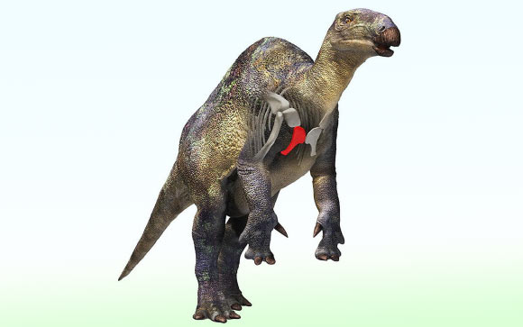
Fossil of Small Duck-Billed Dinosaur Relative Found in Japan
Un fossile d'un petit dinosaure à bec de canard découvert au Japon
os Japon Crétacé fossile Canardia Dinosauria Iguanodontia Styracosterna
Un os fossilisé découvert il y a vingt ans dans le nord-est du Japon appartient à l'un des plus petits dinosaures iguanodontiens jamais découverts dans les roches marines du Crétacé du pays, selon une nouvelle recherche menée par l'Université de Tsukuba. L'article Un fossile d'un petit dinosaure à bec de canard trouvé au Japon est apparu en premier sur Sci.News : Breaking Science News.
20/08/2026 sci-news ⚙ Traduction automatique
1 2 3 4 5 6 7 8 9 10 11 12 13