Galerie d'images
Toutes les images de la base — taxons, formations et intervalles géologiques.
⚠ La fonctionnalité de récupération des images est en cours de test, des images non pertinentes peuvent apparaître.
2,398 image(s)

A, Akmechetosauropus makhkamovi (redrawn from [56]); B, Amblydactylus gethingi (redrawn from [8]); C, Amblydactylus kortmeyeri (redrawn from [57]); D, Apulosauripus federicianus (redrawn from [62]); E, Babatagosauropus bulini (redrawn from [56]); F, Bonaparteichnium tali (redrawn from [66]); G, Brachyguanodonipus prejanensis (redrawn from [68]); H, Camptosaurichnus fasolae (redrawn from [70]); I, Camptosauropus vialovi (redrawn from [74]); J, Caririchnium magnificum (redrawn from [11]); K, Caririchnium leonardii (redrawn from [76]); L-M, Caririchnium protohadrosaurichnos (redrawn from [78]); N, Caririchnium lotus (redrawn from [81]); O, Caririchnium kyoungsookimi (redrawn from [80]); P, Gigantoshiraminesauropus matsuoi (redrawn from [82]); Q, Gypsichnites pacensis (redrawn from [8]); R, Hadrosaurichnoides igeensis (redrawn from [92]); S, Hadrosaurichnus australis (redrawn from [93]); T, Hadrosaurichnus titicaensis (redrawn from [96]); U, Hadrosauripeda hauboldi (redrawn from [98]); V, Hadrosauropodus langstoni (redrawn from [24]); W, Hadrosauropodus nanxiongensis (redrawn from [99]); X, Iguanodonichnus frenkii (redrawn from [70]); Y, Iguanodonipus cuadrupedae (redrawn from [68]).
A, Akmechetosauropus makhkamovi (redrawn from [56]); B, Amblydactylus gethingi (redrawn from [8]); C, Amblydactylus kortmeyeri (redrawn from [57]); D, Apulosauripus federicianus (redrawn from [62]); E, Babatagosauropus bulini (redrawn from [56]); F, Bonaparteichnium tali (redrawn from [66]); G, Brachyguanodonipus prejanensis (redrawn from [68]); H, Camptosaurichnus fasolae (redrawn from [70]); I, Camptosauropus vialovi (redrawn from [74]); J, Caririchnium magnificum (redrawn from [11]); K, Caririchnium leonardii (redrawn from [76]); L-M, Caririchnium protohadrosaurichnos (redrawn from [78]); N, Caririchnium lotus (redrawn from [81]); O, Caririchnium kyoungsookimi (redrawn from [80]); P, Gigantoshiraminesauropus matsuoi (redrawn from [82]); Q, Gypsichnites pacensis (redrawn from [8]); R, Hadrosaurichnoides igeensis (redrawn from [92]); S, Hadrosaurichnus australis (redrawn from [93]); T, Hadrosaurichnus titicaensis (redrawn from [96]); U, Hadrosauripeda hauboldi (redrawn from [98]); V, Hadrosauropodus langstoni (redrawn from [24]); W, Hadrosauropodus nanxiongensis (redrawn from [99]); X, Iguanodonichnus frenkii (redrawn from [70]); Y, Iguanodonipus cuadrupedae (redrawn from [68]).
A, Akmechetosauropus makhkamovi (redrawn from [56]); B, Amblydactylus gethingi (redrawn from [8]); C, Amblydactylus kortmeyeri (redrawn from [57]); D, Apulosauripus federicianus (redrawn from [62]); E, Babatagosauropus bulini (redrawn from [56]); F, Bonaparteichnium tali (redrawn from [66]); G, Brachyguanodonipus prejanensis (redrawn from [68]); H, Camptosaurichnus fasolae (redrawn from [70]); I, Camptosauropus vialovi (redrawn from [74]); J, Caririchnium magnificum (redrawn from [11]); K, Caririchnium leonardii (redrawn from [76]); L-M, Caririchnium protohadrosaurichnos (redrawn from [78]); N, Caririchnium lotus (redrawn from [81]); O, Caririchnium kyoungsookimi (redrawn from [80]); P, Gigantoshiraminesauropus matsuoi (redrawn from [82]); Q, Gypsichnites pacensis (redrawn from [8]); R, Hadrosaurichnoides igeensis (redrawn from [92]); S, Hadrosaurichnus australis (redrawn from [93]); T, Hadrosaurichnus titicaensis (redrawn from [96]); U, Hadrosauripeda hauboldi (redrawn from [98]); V, Hadrosauropodus langstoni (redrawn from [24]); W, Hadrosauropodus nanxiongensis (redrawn from [99]); X, Iguanodonichnus frenkii (redrawn from [70]); Y, Iguanodonipus cuadrupedae (redrawn from [68]).


A badly drew sketch of Brontoholus (a placeholder for when an actual paleoart of Brontotholus is made.

A photograph of partial specimen American Museum of Natural History (AMNH) 22555, posterior dorsal vertebrae, and the sacrum and pelvis (both iliae, and right ischium and pubis) of Anhanguera sp. (formerly often assigned to Anhanguera santanae),[1] from the Early Cretaceous Romualdo Formation (former Romualdo Member of the Santana Formation) of NE Brazil in dorsal view.


Reconstrucuon of Bagaraatan ostromi gen. et sp. n. from the ?mid-Maastrichtian Nemegt Formation of Nemegt, Mongolia.
Shri rapax 3D skeletal reconstruction by Alex Pritchard of Dinosaur Skeletons.

Dorsal vertebra of platecarpus, a cretaceous. Mosasaur from the Niobrara Chalk of Kansas etc. General Collections Keywords: prehistoric archaeology; Paleopathology; Moodie, Roy Lee

Ichthyosaur Skeleton, Platypterygius longmani, in The Museum and Art Gallery of the Northern Territory at Darwin.

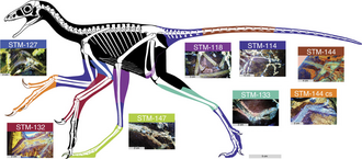
Complete specimen of the Laueropterus vitriolus holotype (LF 6268). The holotype specimen seen under natural light. Major elements are labeled. Abbreviations as follows here and in subsequent figures: ca, caudal vertebra; cp, carpal; cv, cervical vertebrae; dv, dorsal vertebra; fe, femur; hu, humerus; mn, mandible; mt, metatarsal; ph, phalanges; pmm, premaxilla-maxilla; pp, prepubis; pt, pteroid; pv, pelvic plate; r, rib; sa, sacrum; sc, scapulocoracoid; sk, skull part; st, sternum; ta, tarsals; ti, tibia; ul, ulna; un, ungual; wpx, wing phalanges; wmc, wing metacarpal. Scale bar = 100 mm.


Fossil of Pinacosaurus, an ankylosaurian dinosaur Took the photo at Musee d'Histoire Naturelle, Brussels

Cryptarcus ("Chasmosaurus") russelli holotype cranium (CMNFV 8800). (A) Skull in left lateral view (with restored jugal and lower jaw); (B) detail of snout in left lateral view; (C) detail of parietosquamosal frill in dorsal view; (D) detail of supraorbital review in dorsal view; (E) detail of es6 episquamosal; (F) detail of es7 episquamosal. Arrowheads in E and F point to margins of episquamosals.


Chaohusaurus specimen AGM CHS-5, a nearly complete skeleton that is almost as large as AGM I-1. Large scale bars are 10 cm, and short bars 2 cm.

The skull of Janusaurus lundi (PMO 222.654). A: Photo in right lateral view of the skull. B: Right lateral view with interpretation of the individual elements. Abbreviations: a, angular; art, articular; d, dentary; en, external naris; j, jugal; l, lacrimal; mx, maxilla; n, nasal; or, orbit; p, parietal; pmx, premaxilla; po, postorbital; pof, postfrontal; prf, prefrontal; q, quadrate; qj, quadratojugal; sa, surangular; st, supratemporal. Scale = 5 cm.

The holotype and only known specimen of the hauffiopterygian leptonectid, Xiphodracon goldencapensis (ROM VP52596) from Golden Cap, between Charmouth and Seatown, Dorset, UK. The skeleton is exposed in ventrolateral view. The skull has been fully prepared free of matrix whereas most of the skeleton is still in matrix. The left (upper) forefin has been prepared so that it is three-dimensionally preserved and projects upwards. Scale bar represents 20 cm.

Skull of Jinchuanloong niedu (JCMF0132) in left lateral view.


Oneirosaurus caballeroi holotype IGMp879524. A, photograph, drawing and interpretative scheme of the specimen without left mandibular ramus, in right lateral view. B, photograph, drawing and interpretative scheme of the specimen without left mandibular ramus, in left lateral view. Gray fill, sediment; pattern fill, broken bone surface; black, empty space. Scale bars: A = 50 mm; C-D = 30 mm. Abbreviations: an, angular; avc, anterior aperture of the vidian canal; bo, basioccipital; bptm, basipterygoid meniscus; bs, basisphenoid; c, coronoid; d, dentary; ec, ectopterygoid; ep, epipterygoid; ex, exoccipital; f, frontal; im, internal auditory meatus; is, interorbital septum; j, jugal; la, lacrimal; m, mandible; mx, maxilla; na, nasal; op, opisthotic-exoccipital; p, parietal; par, prearticular; pl, palatine; pmx, premaxilla; pof, postorbitofrontal; pr, prootic; prf, prefrontal; ps, parasphenoid; pt, pterygoid; pvc, posterior aperture of the vidian canal q; quadrate; rv, right vomer; sa, surangular; scp, sclerotic plate; smx, septomaxilla; so, supraoccipital; sp, splenial; stp, stapes; VII, IX-XII, exit of cranial nerves.

Life restoration of Afromimus based on skeletal diagram of Masiakasaurus by Scott Hartman.

Holotype of Alcione elainus. Fig. 6 of: Longrich, N. R., Martill, D. M., & Andres, B. (2018). Late Maastrichtian pterosaurs from North Africa and mass extinction of Pterosauria at the Cretaceous-Paleogene boundary. PLoS biology, 16(3), e2001663. --- Original figure legend: A. elainus FSAC-OB 2, holotype partial skeleton and FSAC-OB 217, metacarpal IV. (A) Holotype right humerus in anterior view, (B) holotype right ulna and radius in anterior view, respectively, (C) holotype sternum in left lateral view, (D) referred metacarpal IV, (E) holotype, distal end of left metacarpal IV and left scapulocoracoid, and (F) holotype right femur in posterior view. Abbreviations: co, coracoid; cr, cristospine; dc, distal condyle; dpc, deltopectoral crest; ect, ectepicondyle; fh, femoral head; gl, glenoid; gt, greater trochanter; hh, humeral head; hum, humerus; mcIV, metacarpal IV, pc, proximal cotyle; pf, pneumatic foramen; rad, radius; scpr, supracondylar process; ste, sternum; uln, ulna.

Life restoration of Tawa hallae created by Jeffrey Martz (Petrified Forest National Park, AZ)

Basal cryptoclidid plesiosaur restoration.

Fossil of Quetzalcoatlus, an extinct pterosaur- Took the photo at Senckenberg Museum of Frankfurt


Body mass evolution of Oviraptorosauria. Time calibrated phenograms of Log10 Body Mass (kg) versus time (Ma) for Oviraptorosauria. Blue halos represent 95% confi- dence intervals and branches indicate phylogenetic relation- ships. Each plot displays the same data, but Caenagnathidae is highlighted in green in (A) and Oviraptoridae is highlighted in red in (B) for clarity. Yellow arrows indicate nodes where important changes in body size range occur. Pie charts show ancestral estimations of biogeographic range (as in Fig. 20) for important clades of caenagnathids (A) and oviraptorids (B). Node labels from left to right in (A): Oviraptorosauria; Caenagnathidae; Anomalipes + Caenagnathinae; Caenagnathinae more derived than Apatoraptor pennatus; Anzu + Caenagnathus. Node labels from left to right in (B): Oviraptorosauria; Caenagnathoidea; Oviraptoridae; Heyuanninae (bottom); Citipatinae (top). Colours for node labels as in Fig. 20. Abbreviations: Al, Albian; Ap, Aptian; Ba, Barremian; Be, Berriasian; Ca, Campanian; Ce, Cenomanian; Co, Coniacian; Ha, Hauterivian; Ma, Maastrichtian; S, Santonian; Tu, Turonian; Va, Valanginian.

Dictyoolithidae indet. (eggs in middle), from Lishui, Zhejiang Museum of Natural History (Hangzhou)

Majungasaurus crenatissimus, an abelisaur from the Late Cretaceous of Madagascar, pencil drawing


Representation of the moroccan plioplatecarpinae Khinjaria acuta
Representation of the moroccan plioplatecarpinae Khinjaria acuta

A life reconstruction of the plioplatecarpine Angolasaurus bocagei, alongside the turtle Angolachelys mbaxi. Digital painting.


Eudimorphodon ranzii - photo taken in museums of natural science in Bergamo
Skeletal restoration of Anchiornis huxleyi by Scott Hartman (2017), incorporating the soft tissue outlines revealed by laser fluorescence studies.

Lithography of specimen ANSP 9534, the lectotype of Deinodon horridus.


Left postorbital horncore of ‘Ceratops montanus’ (USNM 2411) in ventrolateral view.


PRESERVED_SPECIMEN; ; ; microslide; HOLOTYPE. See: Sanders, Howard L. 1955. The Cephalocarida, a new subclass of Crustacea from Long Island Sound. Proceedings of the National Academy of Sciences. 41 (1): 61-66.; CSBR Slide Grant Image 2015; IZ number 3617; lot count 1; Microslide 01, balsam, whole mount; 1954-07-23T00:00:00Z


Brontosaurus excelsus in the Yale Peabody Museum of Natural History. Brontosaurus excelsus Inv no. YPM 1980 (holotype specimen of the species) Discoverer William H. Reed 1879 Locality Como Bluff, Wyoming Age Morrison Formatian, Jurassic period, 150 million years ago

Bothriospondylus suffossus. Fig. 1. Hind view of terminal centrum of sacral vertebra. Fig. 2. Right side view of the same. Fig. 3. hæmal view of the same. Fig. 4. Neural view of mutilated centrum of sacral vertebra, restored in outline. Fig. 5. Right side view of the same. Fig. 6. hæmal view of the same, restored in outline. All the figures are of the natural size. From the Kimmeridge Clay at Swindon, Wilts. In the British Museum.

Tastavinsaurus, pencil drawing, digital coloring

Photo montage of several representatives members of the clade Dracohors (dinosaurs and their extinct relatives): Asilisaurus Borealopelta Triceratops Giganotosaurus

Identifier: waterreptilesofp1914will Title: Water reptiles of the past and present Year: 1914 (1910s) Authors: Williston, Samuel Wendell, 1851-1918 Subjects: Aquatic reptiles Publisher: Chicago, Ill., The University of Chicago Press Contributing Library: Boston Public Library Digitizing Sponsor: Boston Public Library View Book Page: Book Viewer About This Book: Catalog Entry View All Images: All Images From Book Click here to view book online to see this illustration in context in a browseable online version of this book. Text Appearing Before Image: served and very complete skeletons ofdifferent species of ichthyosaurs from the Jurassic deposits ofWiirtemberg, in which remains of these animals occur in great 112 WATER REPTILES OF THE PAST AND PRESENT profusion. His researches, and those of several authors since then,supplementing and confirming or disproving those of the manyobservers made during the preceding seventy years, have finallydetermined almost perfectly the complete structure of the moretypical ichthyosaurs, enabling us to infer not a little as to theirhabits and distribution in the old Jurassic oceans. Within thepast few years the discoveries of Professor J. C. Merriam of Cali-fornia have likewise added greatly to our knowledge of the earlierichthyosaurs. It may now truthfully be said that of no group ofextinct reptiles do we have a more complete and satisfactory knowl-edge than of the ichthyosaurs. Nevertheless we have yet very much more to learn about theorder Ichthyosauria as a whole—whence they came and how they Text Appearing After Image: Fig. 52.—Ichthyosaurus quadricissus.museum, from Dr. Dreverman. Photograph of specimen in Senckenberg originated; what their nearest kin were among other reptiles; andespecially, more about the connecting links between them andterrestrial reptiles. They have, as an order, so isolated a position,are so widely separated from all other reptiles in structure, that theyhave long been a puzzle to paleontologists. Like the whales andother cetaceans among mammals, we know the ichthyosaurs wellin the plenitude of their power and the fulness of their development,but have yet only an imperfect knowledge of their earlier history,and none whatever of their earliest. However, as will be seenfarther on, the recent discoveries by Merriam have shed much lighton some of the stages of their evolution. So nearly perfectly wereall the later ichthyosaurs adapted to their life in the water that itwas believed by nearly all paleontologists until about a score of years ICHTHYOSAURIA 3 ago that they had desc Note About Images Please note that these images are extracted from scanned page images that may have been digitally enhanced for readability - coloration and appearance of these illustrations may not perfectly resemble the original work.


Skeletal restoration of Preondactylus bufarini.
Skeletal restoration of Preondactylus bufarini.

Jeholornis prima skeleton (IVPP V13350) on display at the Paleozoological Museum of China.
Jeholornis prima skeleton (IVPP V13350) on display at the Paleozoological Museum of China.

Model of vulcanodon in JuraPark, Solec Kujawski, Poland
Model of vulcanodon in JuraPark, Solec Kujawski, Poland


Alioramus altai skull in the exhibit, T. rex, The Ultimate Predator, in the American Museum of Natural History (with permission by Ben Miller).