Galerie d'images
Toutes les images de la base — taxons, formations et intervalles géologiques.
⚠ La fonctionnalité de récupération des images est en cours de test, des images non pertinentes peuvent apparaître.
2,347 image(s)


A, Four consecutive track casts of Minisauripus isp.; B, Natural impression slab of second left and right feet tracks; C, Isolated fifth track of Minisauripus isp.


fossil of Prenocephale prenes, an extinct ornithischian

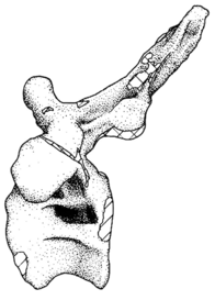
AMNH 0044, Sphaerotholus buchholtzae, in dorsal view with arrows denoting dorsal lesions


Figure 24: Reconstruction of Kwanasaurus williamparkeri. (A) Skeletal reconstruction with elements based on individuals of varied sizes, all scaled under the assumption that Kwanasaurus is proportioned similarly to Silesaurus. (B) Life reconstruction. Scale bars = 10 cm, given for probable largest specimen (DMNH EPV.34579) and one of the smallest specimens (DMNH EPV.63139).

Dentary of Echinodon becklesii from the Lower Cretaceous Purbeck Formation of England. Left dentary (NHMUK 48215b) in lateral (A), medial (B), and dorsal (C) views.
Dentary of Echinodon becklesii from the Lower Cretaceous Purbeck Formation of England. Left dentary (NHMUK 48215b) in lateral (A), medial (B), and dorsal (C) views.

Comparison of cranial features between closely related southern Laramidian taxa; (A), Akainacephalus johnsoni (UMNH VP 20202) from the Late Cretaceous Kaiparowits Formation of Utah; and (B), Nodocephalosaurus kirtlandensis (SMP VP-900) from the Late Cretaceous Kirtland Formation of New Mexico, in left lateral views. Various synapomorphies are shared with N. kirtlandensis (highlighted in black and white arrows) and includes “flaring nostrils”; enlarged, laterally projecting, loreal osteoderms that are situated directly dorsal to the external nares. Other synapomorphies include pyramid-shaped nasal and frontal osteoderms positioned on the dorsal regions of the skull. A number of significant differences have been observed between both specimens; in A. johnsoni, the anterior, and posterior supraorbital bosses form an enlarged element that is somewhat backswept, whereas in N. kirtlandensis, the posterior and anterior supraorbital bosses are clearly defined as individual osteoderms, and are much smaller in size. Additionally, the squamosal horn in Akainacephalus is very small but is prominent and tetrahedrally shaped in Nodocephalosaurus. The quadratojugal horn in Akainacephalus is massive, has a subtriangular morphology in lateral view and projects almost entirely ventral, whereas in Nodocephalosaurus, the quadratojugal horn is smaller and has a typical fin-shaped morphology. Study sites: asob, anterior supraorbital boss; ext naris, external naris; laca, lacrimal caputegulum; loca, loreal caputegulum; naca, nasal caputegulae; orb, orbit; psob, posterior supraorbital boss; qjh, quadratojugal horn; sqh, squamosal horn.


Diamantinasaurus matildae gen. et sp. nov. (AODF 603) A. Right side B. Left side (both silhouettes with sketched in bone parts of the material currently known at publishing date; scale bars: 5 x 5 = 25 m; complemented with height data here)


A life reconstruction of the titanosaur Aeolosaurus rionegrinus in lateral view. The neck and head reconstructed based on its relative Rapetosaurus; the curvature of the tail is based on da Silva Vidal et al. 2020 (doi: 10.1080/08912963.2020.1745791)


Fossil skeleton of Qianichthyosaurus zhoui on display at the Geological Museum of China.

Photograph of a fossil cast of a Keichousaurus hui skeleton taken at the North American Museum of Ancient Life.


Skull of Pistosaurus longaevus (cast - USNM 16107) in right, left and posterior views.
Skull of Pistosaurus longaevus (cast - USNM 16107) in right, left and posterior views.


Tooth of cf. Zapsalis, with close up of denticles. Specimen UALVP 49582 from the Milk River Formation.


Caenagnathasia martinsoni Currie, Godfrey & Nessov, 1994. Mandibles of the holotype CMGP 401/12457 (left) and paratype CMGP 402/12457 (right). Mandibles in dorsal view, ventral view, and right lateral view. Paratype is copied, flipped and paired with the original on the right side.

Saurexallopus, a four toed dinosaur or bird trace fossil (jr syn Exallopus

Skull replica of Vagaceratops at Canadian Museum of Nature

Skull cast of Anchiceratops ornatus (original specimen: TMP 1983.001.0001) on display at the University of Michigan Museum of Natural History.


Pararhabdodon isonensis, maxillae. A. Right maxilla (IPS 36327) in lateral view. B. Medial view of same. C. Left maxilla (IPS 693-6) in medial view. D. Lateral view of same.


* Wintonotitan wattsi gen. et sp. nov. (QMF 7292) (Silhouette with sketched in bone parts of the material currently known at publishing date; scale bar: size unknown — not mentioned in original source)


Digital illustration of the Sauropod Dinosaur Isisaurus colberti from the Late Cretaceous (Maastrichtian) of India (72.2-66 MYA). References include Jain & Bandyopadhyay (1997), several papers from Wilson et al. and skeletal reconstruction by Scott Hartman. Illustrated by Ansh Saxena. About Isisaurus– Isisaurus colberti (=Titanosaurus colberti) was a species of Titanosaurian Sauropod Dinosaur from the Late Cretaceous (Maastrichtian) age in the Indian Subcontinent. Isisaurus lived sympatrically with another Titanosaurian Sauropod Jainosaurus. It also lived alongside Theropods like Rajasaurus, Rahiolisaurus, Indosuchus etc. Remains of Isisaurus come from the Lameta formation of Central India.

Holotype material of Aegyptosaurus, based on plates in Stromer 1932. Scale bar = 1 meter


Cranial remains of Arthropterygius chrisorum CCMGE 17–44/13328 (A–J) and PMO 222.669 (L, M). (A, B) Right postfrontal in ventral (A) and dorsal (B) views. (C) Left lateral view on articulated postfrontal, prefrontal and nasal. (D) Left prefrontal in ventral view. (E, F) Right prefrontal in ventral (E) and dorsal (F) views. (G, H) left nasal in dorsal (G) and ventral (H) views. (I, J) Left jugal in medial (I) and lateral (J) views. (K) Cranial reconstruction, showing the depicted elements (colored). (L, M) oblique dorsal view and interpretation of sutures of the skull roof of PMO 222.669. Abbreviations: ffr, facet for the frontal; fnas, facet of the nasal; fpo, facet for the postorbital; fpref, facet for the prefrontal; fqj, facet for the quadratojugal; fsut, facet for the supratemporal; lw, lateral wing of the nasal lamella; nas, nasal; par, parietal; pf, parietal foramen; pref, prefrontal; sut, supratemporal. Both scale bars represent 10 cm.


Holotype AMNH 6516. Birdlike reptile Visit my blog at ideonexus.com
Holotype AMNH 6516. Birdlike reptile Visit my blog at ideonexus.com

Skeletal reconstructions of Dinosaur Park Formation caenagnathids. Skeletal reconstructions of Citipes elegans (left), Chirostenotes pergracilis (middle), and Caenagnathus collinsi (right), showing variation in skeletal representation and body size. Previously referred material is indicated in white and newly referred material is indicated in red for each taxon. Blue asterisks indicate elements that have been histologically sampled for each taxon. Citipes elegans: dentaries, metatarsal IV; Chirostenotes pergracilis: dentaries, tibia; Caenagnathus collinsi: pubis.


Megapnosaurus is a coelophysid theropod dinosaur from the Early Jurassic Period of Africa. It was a lightly built bipedal carnivore that grew to just over 2 m long and 13 kg in body mass. Its close relation to Coelophysis has caused some confusion in classifying the genus - it had a slender build and curved S-shaped neck, but was more robust. Comparisons between the scleral rings of Megapnosaurus and modern birds and reptiles indicate that it may have been nocturnal.

Fossil of Leptoceratops at the Canadian Museum of Nature, Ottawa

Restoration of Asiaceratops salsopaludalis a ceratopsian dinosaur from the Late Cretaceous of Uzbekistan

MSNVE 3714, Ouranosaurus nigeriensis. The mounted specimen as exhibited today at the MSNVE. For scale, the right femur is 920 mm long.
