Image gallery
All images in the database — taxa, formations and geological intervals.
⚠ The image retrieval feature is currently being tested — irrelevant images may appear.
2,347 image(s)

Lower Triassic fossil footprint (ichnite) of the ichnogenus Chirotherium, probably caused by an early archosaur, and first discovered 1833 in Hildburghausen (Thuringia, Germany). This specimen, however, ist from the Helsby Sandstone of the Storeton Quarry near Liverpool. Its species name is Chirotherium storetonense.[1]
Lower Triassic fossil footprint (ichnite) of the ichnogenus Chirotherium, probably caused by an early archosaur, and first discovered 1833 in Hildburghausen (Thuringia, Germany). This specimen, however, ist from the Helsby Sandstone of the Storeton Quarry near Liverpool. Its species name is Chirotherium storetonense.[1]
Lower Triassic fossil footprint (ichnite) of the ichnogenus Chirotherium, probably caused by an early archosaur, and first discovered 1833 in Hildburghausen (Thuringia, Germany). This specimen, however, ist from the Helsby Sandstone of the Storeton Quarry near Liverpool. Its species name is Chirotherium storetonense.[1]
Lower Triassic fossil footprint (ichnite) of the ichnogenus Chirotherium, probably caused by an early archosaur, and first discovered 1833 in Hildburghausen (Thuringia, Germany). This specimen, however, ist from the Helsby Sandstone of the Storeton Quarry near Liverpool. Its species name is Chirotherium storetonense.[1]
Lower Triassic fossil footprint (ichnite) of the ichnogenus Chirotherium, probably caused by an early archosaur, and first discovered 1833 in Hildburghausen (Thuringia, Germany). This specimen, however, ist from the Helsby Sandstone of the Storeton Quarry near Liverpool. Its species name is Chirotherium storetonense.[1]
Lower Triassic fossil footprint (ichnite) of the ichnogenus Chirotherium, probably caused by an early archosaur, and first discovered 1833 in Hildburghausen (Thuringia, Germany). This specimen, however, ist from the Helsby Sandstone of the Storeton Quarry near Liverpool. Its species name is Chirotherium storetonense.[1]

Argentinosaurus huinculensis reconstruction at Museo Municipal Carmen Funes, Plaza Huincul, Neuquén, Argentina.


Left ilium of the camarasauromorph sauropod Brontomerus mcintoshi from the Lower Cretaceous Cedar Mountain Formation of Utah, type specimen OMNH 66430 in lateral view reconstructed from the three fragments (A), and ventral view (B).


Lusotitan atalaiensis. Photographs of right humerus (proximal half) in (A) anterior (slightly oblique as a result of mounted position), (B) medial, (C) proximal, (D) lateral, and (E) posterior views. Abbreviations: dtp, deltopectoral crest; hh, humeral head. Scale bar = 200 mm.

Middle cervical vertebra of Vouivria damparisensis (MNHN.F.1934.6 DAM 6). (A) Left lateral view, (B) anterior view, (C) right lateral view; (D) posterior view. Abbreviations: cpol, centropostzygapophyseal lamina; di, diapophysis; cprl, centroprezygapophyseal lamina; espol, expanded spinopostzygapophyseal lamina; no, notch; ns, notch; pa, parapophysis; pn, pneumatic foramen; pocdf, postzygapophyseal centrodiapophyseal fossa; podl, postzygodiapophyseal lamina; poz, postzygapophysis; prz, prezygapophysis; ri, ridge; sdf, spinodiapophyseal fossa; spol, spinopostzygapophyseal lamina; sprl, spinoprezygapophyseal lamina. Scale bar equals 10 cm.


Recreación de Galvesaurus, posible saurópodo macronario basal de la Península Ibérica


Liaoningotitan sinensis, mount, Liaoning Palaeontological Museum


fossil Europasaurus, Aathal Dinosaur Museum.

Skeleton of Tehuelchesaurus at the Museo Paleontológico Egidio Feruglio in Trelew, Argentina


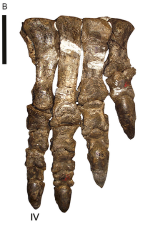
Rhoetosaurus brownei (QM F1659; holotype [part]) right crus and pes in anterodorsal view. Scale = 20 cm.

Reconstructed skull of the turiasaurian Mierasaurus, based on the holotype UMNH.VP.26004.


Left Femur in caudal view Lapparentosaurus madagascariensis missing the mid-shaft section; FC = fibular condyle; FH = femoral head; GT = greater trochanter; ICG = intercondylar groove; TC = tibial condyle. Scale = 10 cm.

Fig. 2. Side view of crown of tooth of Cardiodon rugulosus. Fig. 3. Fore end of the same tooth. Fig. 4. Hind end of the crown of another tooth of Cardiodon rugulosus. Fig. 5. Magnified view of markings on the surface of the enamel of the same tooth. All the figures are of the natural size. 2-5 are from the Forest Marble of Bradford, Wilts. In the Collection of Channing Pearce, Esq., of that town.

Left humerus of Haestasaurus becklesii (NHMUK R1870). A, anterior view; B, posterior view; Abbreviations: af, anconeal fossa; dp, deltopectoral crest; hh, humeral head; ltf, lateral triceps fossa; mtf, medial triceps fossa. This fossil was found in strata of the Hastings Beds (late Berriasian—Valanginian in age) on the coast near Hastings, East Sussex, England.

Fig. 1. Type material of Amygdalodon patagonicus Cabrera. 1947. A. B. Tooth, MLP 46-VIII-21-1/13, in lingual (A) and labial (B) view. C. D, E. Cervical vertebra, MLP 46-VIII-21-1/8. in right lateral (C, stereopair), ventral (D) and left lateral (E) view. F, G. Right cervical prezygapophysis, MLP 46-VIII-21-117. in dorsal (F) and medial (G) view. H. Anterior dorsal neural spine, MLP 46-VIII-21-116. in posterior view. I, J, K. Posterior dorsal vertebra, MLP 46-VIII-21-112, lectotype, in right lateral view (I, stereopair), right lateral view, with right wall of neural canal remived (J), and posterior (K) view. L, M. Caudal vertebra, MLP 46-VIII-21-1/3, in left lateral (L) and posterior (M) view. N. Caudal vertebra, MLP 46-VIII-21-1/4, in right lateral view. (O). Proximal fragment of a dorsal rib, MLP 46-VIII-21-1/9, in anterolateral view. Abbreviations: acdl, anterior centrodiapophyseal lamina; cprl, centroprezygapophyseal lamina; g, groove; k, ventral keel; pcdl, posterior centrodiapophyseal lamina; pod, postzygodiapophyseal lamina; poz, postzygapophysis; pp, parapophysis; prdl, prezygodiapophyseal lamina; spol, spinopostzgapophyseal lamina; sprl, spinoprezygapophyseal lamina; tprl, intraprezygapophyseal lamina. Scale bars 1 cm (A, B) and 10 cm (C-0).

Exhibit in the Naturhistorisches Museum, Braunschweig, Germany.


Skull diagram showing the known material of Aardonyx. Based on photographs and measurements in original description and supplementary material. Scale bar = 10 cm


Right hind limb of the sauropodomorph dinosaur Musankwa sanyatiensis gen. et sp. nov. (NHMZ 2521) from the Pebbly Arkose Formation (Norian, Upper Triassic) of Spurwing Island, Zimbabwe. A. Right femur in posterior (A1), lateral (A2), anterior (A3), medial (A4), proximal (A5), and distal (A6) views. B. Right tibia with conjoined astragalus in anterior (B1), lateral (B2), posterior (B3), medial (B4), and proximal (B5) views.

Reconstruction of Tuebingosaurus maierfritzorum gen. et sp. nov. as a quadruped dinosaur, using the outline of Riojasaurus as a base ‒ next to the silhouette of Friedrich von Huene. The drawing of the bones is based on and modified from the original illustrations of specimen “GPIT IV” in von Huene (1932, pl. 38) that have been replicated in the literature. The right fibula is marked in grey as it was found nearby with similar measurements to the left fibula and has been assumed to be part of the same individual.


Skull of the new basal sauropodomorph Leyesaurus marayensis (PVSJ 706). Photograph of the skull (A) and interpretative drawing (B) in lateral view. Dark grey color represents matrix and light grey color represents foraminae. Abbreviations: a, angular; aoF, antorbital fenestra; aoFo; antorbital fossa; Apmx, ascending process of the maxilla; d, dentary; f, frontal; itF, infratemporal fenestra; j, jugal; l, lacrimal; laoFo; lacrimal antorbital fossa; mF, mandibular fenestra; mx, maxilla; n, nasal; O, orbit; p, parietal; pf, prefrontal; pm, premaxilla; po, postorbital; rug, platform-like rugosities; q, quadrate; qj, quadratejugal; Rmx, ridge of the ascending process of the maxilla; sa, surangular; snf, subnarial foramen. Scale bar equals 1cm.

Holotype fossils of Glacialisaurus (PR 1823); metatarsals, tibia, fibula, ankle bones

Left maxilla of the silesaurid Agnosphitys cromhallensis from the Late Triassic (Rhaetian) of England.


Amniote humerus originally referred to Actiosaurus in anterior view (2). Figures 1 and 2 after Sauvage (1883).


Pantydraco caducus, a sauropodomorph from the Late Triassic or Early Jurassic of England, after Yates, 2003, pencil drawing, digital coloring
(B) Kholumolumo ellenbergerorum (NMQR1705). Reverse image in anterior view from Krupandan (2019).

3D Skeletal reconstruction of Huayracursor jaguensis, with known remains colored in orange


Digital drawing of the known skeletal remains of Nhandumirim waldsangae. Known elements represented in white and unknown in gray. This is an alternative version, reconstructing the elements based on early sauropodomorphs.