Galerie d'images
Toutes les images de la base — taxons, formations et intervalles géologiques.
⚠ La fonctionnalité de récupération des images est en cours de test, des images non pertinentes peuvent apparaître.
4,274 image(s)

Middle cervical vertebra of Vouivria damparisensis (MNHN.F.1934.6 DAM 6). (A) Left lateral view, (B) anterior view, (C) right lateral view; (D) posterior view. Abbreviations: cpol, centropostzygapophyseal lamina; di, diapophysis; cprl, centroprezygapophyseal lamina; espol, expanded spinopostzygapophyseal lamina; no, notch; ns, notch; pa, parapophysis; pn, pneumatic foramen; pocdf, postzygapophyseal centrodiapophyseal fossa; podl, postzygodiapophyseal lamina; poz, postzygapophysis; prz, prezygapophysis; ri, ridge; sdf, spinodiapophyseal fossa; spol, spinopostzygapophyseal lamina; sprl, spinoprezygapophyseal lamina. Scale bar equals 10 cm.


Recreación de Galvesaurus, posible saurópodo macronario basal de la Península Ibérica
Recreación de Galvesaurus, posible saurópodo macronario basal de la Península Ibérica


Liaoningotitan sinensis, mount, Liaoning Palaeontological Museum


fossil Europasaurus, Aathal Dinosaur Museum.

Skeleton of Tehuelchesaurus at the Museo Paleontológico Egidio Feruglio in Trelew, Argentina


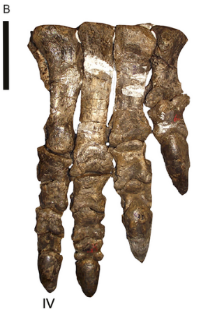
Rhoetosaurus brownei (QM F1659; holotype [part]) right crus and pes in anterodorsal view. Scale = 20 cm.

Reconstructed skull of the turiasaurian Mierasaurus, based on the holotype UMNH.VP.26004.


Left Femur in caudal view Lapparentosaurus madagascariensis missing the mid-shaft section; FC = fibular condyle; FH = femoral head; GT = greater trochanter; ICG = intercondylar groove; TC = tibial condyle. Scale = 10 cm.

Fig. 2. Side view of crown of tooth of Cardiodon rugulosus. Fig. 3. Fore end of the same tooth. Fig. 4. Hind end of the crown of another tooth of Cardiodon rugulosus. Fig. 5. Magnified view of markings on the surface of the enamel of the same tooth. All the figures are of the natural size. 2-5 are from the Forest Marble of Bradford, Wilts. In the Collection of Channing Pearce, Esq., of that town.

Left humerus of Haestasaurus becklesii (NHMUK R1870). A, anterior view; B, posterior view; Abbreviations: af, anconeal fossa; dp, deltopectoral crest; hh, humeral head; ltf, lateral triceps fossa; mtf, medial triceps fossa. This fossil was found in strata of the Hastings Beds (late Berriasian—Valanginian in age) on the coast near Hastings, East Sussex, England.

Fig. 1. Type material of Amygdalodon patagonicus Cabrera. 1947. A. B. Tooth, MLP 46-VIII-21-1/13, in lingual (A) and labial (B) view. C. D, E. Cervical vertebra, MLP 46-VIII-21-1/8. in right lateral (C, stereopair), ventral (D) and left lateral (E) view. F, G. Right cervical prezygapophysis, MLP 46-VIII-21-117. in dorsal (F) and medial (G) view. H. Anterior dorsal neural spine, MLP 46-VIII-21-116. in posterior view. I, J, K. Posterior dorsal vertebra, MLP 46-VIII-21-112, lectotype, in right lateral view (I, stereopair), right lateral view, with right wall of neural canal remived (J), and posterior (K) view. L, M. Caudal vertebra, MLP 46-VIII-21-1/3, in left lateral (L) and posterior (M) view. N. Caudal vertebra, MLP 46-VIII-21-1/4, in right lateral view. (O). Proximal fragment of a dorsal rib, MLP 46-VIII-21-1/9, in anterolateral view. Abbreviations: acdl, anterior centrodiapophyseal lamina; cprl, centroprezygapophyseal lamina; g, groove; k, ventral keel; pcdl, posterior centrodiapophyseal lamina; pod, postzygodiapophyseal lamina; poz, postzygapophysis; pp, parapophysis; prdl, prezygodiapophyseal lamina; spol, spinopostzgapophyseal lamina; sprl, spinoprezygapophyseal lamina; tprl, intraprezygapophyseal lamina. Scale bars 1 cm (A, B) and 10 cm (C-0).

Exhibit in the Naturhistorisches Museum, Braunschweig, Germany.


Skull diagram showing the known material of Aardonyx. Based on photographs and measurements in original description and supplementary material. Scale bar = 10 cm


Right hind limb of the sauropodomorph dinosaur Musankwa sanyatiensis gen. et sp. nov. (NHMZ 2521) from the Pebbly Arkose Formation (Norian, Upper Triassic) of Spurwing Island, Zimbabwe. A. Right femur in posterior (A1), lateral (A2), anterior (A3), medial (A4), proximal (A5), and distal (A6) views. B. Right tibia with conjoined astragalus in anterior (B1), lateral (B2), posterior (B3), medial (B4), and proximal (B5) views.

Reconstruction of Tuebingosaurus maierfritzorum gen. et sp. nov. as a quadruped dinosaur, using the outline of Riojasaurus as a base ‒ next to the silhouette of Friedrich von Huene. The drawing of the bones is based on and modified from the original illustrations of specimen “GPIT IV” in von Huene (1932, pl. 38) that have been replicated in the literature. The right fibula is marked in grey as it was found nearby with similar measurements to the left fibula and has been assumed to be part of the same individual.


Skull of the new basal sauropodomorph Leyesaurus marayensis (PVSJ 706). Photograph of the skull (A) and interpretative drawing (B) in lateral view. Dark grey color represents matrix and light grey color represents foraminae. Abbreviations: a, angular; aoF, antorbital fenestra; aoFo; antorbital fossa; Apmx, ascending process of the maxilla; d, dentary; f, frontal; itF, infratemporal fenestra; j, jugal; l, lacrimal; laoFo; lacrimal antorbital fossa; mF, mandibular fenestra; mx, maxilla; n, nasal; O, orbit; p, parietal; pf, prefrontal; pm, premaxilla; po, postorbital; rug, platform-like rugosities; q, quadrate; qj, quadratejugal; Rmx, ridge of the ascending process of the maxilla; sa, surangular; snf, subnarial foramen. Scale bar equals 1cm.

Holotype fossils of Glacialisaurus (PR 1823); metatarsals, tibia, fibula, ankle bones

Left maxilla of the silesaurid Agnosphitys cromhallensis from the Late Triassic (Rhaetian) of England.

Amniote humerus originally referred to Actiosaurus in anterior view (2). Figures 1 and 2 after Sauvage (1883).


Pantydraco caducus, a sauropodomorph from the Late Triassic or Early Jurassic of England, after Yates, 2003, pencil drawing, digital coloring
(B) Kholumolumo ellenbergerorum (NMQR1705). Reverse image in anterior view from Krupandan (2019).

3D Skeletal reconstruction of Huayracursor jaguensis, with known remains colored in orange


Digital drawing of the known skeletal remains of Nhandumirim waldsangae. Known elements represented in white and unknown in gray. This is an alternative version, reconstructing the elements based on early sauropodomorphs.


Three-dimensional skull of BMT 1955.G35.1, Protoichthyosaurus prostaxalis. (B) Skull in left lateral view, as reconstructed in 2015. (C) Skull in right lateral view, as reconstructed in 2015. Note the distinctive asymmetric maxilla with long, narrow anterior process. Teeth are not in their original positions. Scale bar represents 20 cm.

Figure description from paper: "A Cast of BES SC 999, the holotype of Besanosaurus leptorhynchus and (B) interpretative drawing (modified from Dal Sasso & Pinna, 1996). Foetal remains are highlighted with green lines. a astragalus, c calcaneum, Cl clavicle, Co coracoid, Fe femur, Fi Fibula, H humerus, i intermedium, Il Ilium, Is Ischium, P pubis, p pisiform, R radius, r radiale, S scapula, T Tibia, U Ulna, u ulnare; 2, 3, and 4, distal carpals and tarsals; II, III, IV, and V, metacarpals and metatarsals. The apostrophe (‘) indicates left elements. Scale bar represents 50 cm"


Exhibit in the Royal Ontario Museum, Toronto, Ontario, Canada. This exhibit is old enough so that it is in the public domain, and photography was permitted in the museum. I took this photograph and release it into the public domain.


A figure from Notes on Osteology of Baptanodon. With a Description of a New Species.


The holotype of Dianmeisaurus mutaensis (HFUT MT-21-08-001). (A) the skeleton in dorsal view; (B) the counterpart of (A) (natural mold). Scale bars equal 1 cm.

Skeleton of WGSC SPC V 1304 (juvenile Brevicaudosaurus jiyangshanensis). A, photograph of the specimen in dorsal view. B, interpretive drawing.

Uncovered skeleton of Brachauchenius sp. VL from the late Barremian Formacio´n Paja of Loma La Cabrera, Villa de Leyva area, in dorsal view. The far posterior part (ischia, tail) and the right hind limb are not preserved.

Skull of Megacephalosaurus eulerti FHSM VP-321 in dorsal and palatal view. Right mandible in medial and lateral views.


