←

écaille

Partie anatomique

217 image(s) · 21 Actualités

Galerie d'images

Skeletal reconstruction of Eotyrannus lengi IWCMS: 1997.550. New skeletal reconstruction of Eotyrannus lengi, depicting only those elements preserved in the holotype. The positions shown for some of the isolated vertebrae and ribs are conjectural. Scale bar: 100 cm.
Taxons Eotyrannus

Skeletal reconstruction of Eotyrannus lengi IWCMS: 1997.550. New skeletal reconstruction of Eotyrannus lengi, depicting only those elements preserved in the holotype. The positions shown for some of the isolated vertebrae and ribs are conjectural. Scale bar: 100 cm.

écaille holotype Eotyrannus
Diagram featuring the holotype skull (ZPAL MgD-I/105) of Tylocephale gilmorei in left lateral view. Scale bar equals 5 cm (50 mm). Based on Maryańska & Osmólska, 1974,[1] and Sullivan, 2006.[2]
References

↑ (1974). "Pachycephalosauria, a new suborder of ornithischian dinosaurs". Palaeontologica Polonica (30): 45−102.

↑ (2006). "A taxonomic review of the Pachycephalosauridae (Dinosauria: Ornithischia)". New Mexico Museum of Natural History and Science Bulletin (35): 347–365.
Taxons Tylocephale

Diagram featuring the holotype skull (ZPAL MgD-I/105) of Tylocephale gilmorei in left lateral view. Scale bar equals 5 cm (50 mm). Based on Maryańska & Osmólska, 1974,[1] and Sullivan, 2006.[2] References ↑ (1974). "Pachycephalosauria, a new suborder of ornithischian dinosaurs". Palaeontologica Polonica (30): 45−102. ↑ (2006). "A taxonomic review of the Pachycephalosauridae (Dinosauria: Ornithischia)". New Mexico Museum of Natural History and Science Bulletin (35): 347–365.

écaille musée Mexique holotype +6
An overview of the known fossil material of Tyrannomimus fukuiensis. Fossil specimens shown in are not in the same scale. The bar is scaled for the paratype (FPDM-V-10295). The holotype (FPDM-V-11333) and paratype in are colored in red and blue, respectively. The right femur in is mirrored from the left one (FPDM-V-11338).
Taxons Tyrannomimus

An overview of the known fossil material of Tyrannomimus fukuiensis. Fossil specimens shown in are not in the same scale. The bar is scaled for the paratype (FPDM-V-10295). The holotype (FPDM-V-11333) and paratype in are colored in red and blue, respectively. The right femur in is mirrored from the left one (FPDM-V-11338).

écaille fossile holotype spécimen +1
Fig. 1. Type material of Amygdalodon patagonicus Cabrera. 1947. A. B. Tooth, MLP 46-VIII-21-1/13, in lingual (A) and labial (B) view. C. D, E. Cervical vertebra, MLP 46-VIII-21-1/8. in right lateral (C, stereopair), ventral (D) and left lateral (E) view. F, G. Right cervical prezygapophysis, MLP 46-VIII-21-117. in dorsal (F) and medial (G) view. H. Anterior dorsal neural spine, MLP 46-VIII-21-116. in posterior view. I, J, K. Posterior dorsal vertebra, MLP 46-VIII-21-112, lectotype, in right lateral view (I, stereopair), right lateral view, with right wall of neural canal remived (J), and posterior (K) view. L, M. Caudal vertebra, MLP 46-VIII-21-1/3, in left lateral (L) and posterior (M) view. N. Caudal vertebra, MLP 46-VIII-21-1/4, in right lateral view. (O). Proximal fragment of a dorsal rib, MLP 46-VIII-21-1/9, in anterolateral view. Abbreviations: acdl, anterior centrodiapophyseal lamina; cprl, centroprezygapophyseal lamina; g, groove; k, ventral keel; pcdl, posterior centrodiapophyseal lamina; pod, postzygodiapophyseal lamina; poz, postzygapophysis; pp, parapophysis; prdl, prezygodiapophyseal lamina; spol, spinopostzgapophyseal lamina; sprl, spinoprezygapophyseal lamina; tprl, intraprezygapophyseal lamina. Scale bars 1 cm (A, B) and 10 cm (C-0).
Taxons Amygdalodon

Fig. 1. Type material of Amygdalodon patagonicus Cabrera. 1947. A. B. Tooth, MLP 46-VIII-21-1/13, in lingual (A) and labial (B) view. C. D, E. Cervical vertebra, MLP 46-VIII-21-1/8. in right lateral (C, stereopair), ventral (D) and left lateral (E) view. F, G. Right cervical prezygapophysis, MLP 46-VIII-21-117. in dorsal (F) and medial (G) view. H. Anterior dorsal neural spine, MLP 46-VIII-21-116. in posterior view. I, J, K. Posterior dorsal vertebra, MLP 46-VIII-21-112, lectotype, in right lateral view (I, stereopair), right lateral view, with right wall of neural canal remived (J), and posterior (K) view. L, M. Caudal vertebra, MLP 46-VIII-21-1/3, in left lateral (L) and posterior (M) view. N. Caudal vertebra, MLP 46-VIII-21-1/4, in right lateral view. (O). Proximal fragment of a dorsal rib, MLP 46-VIII-21-1/9, in anterolateral view. Abbreviations: acdl, anterior centrodiapophyseal lamina; cprl, centroprezygapophyseal lamina; g, groove; k, ventral keel; pcdl, posterior centrodiapophyseal lamina; pod, postzygodiapophyseal lamina; poz, postzygapophysis; pp, parapophysis; prdl, prezygodiapophyseal lamina; spol, spinopostzgapophyseal lamina; sprl, spinoprezygapophyseal lamina; tprl, intraprezygapophyseal lamina. Scale bars 1 cm (A, B) and 10 cm (C-0).

écaille dent vertèbre Amygdalodon
Selected elements used in the diagnosis of Saltriovenator zanellai n. gen. n. sp.

Right humerus in medial (A), frontal (B) and distal (C) views; (D) left scapula, medial view; (E) right scapular glenoid and coracoid, lateral view; (F) furcula, ventral view; tooth, labial (G) and apical (H) views; (I) left humerus, medial view; right second metacarpal in dorsal (J), lateral (L) and distal (N) views; first phalanx of the right second digit in dorsal (K), lateral (M) and proximal (O) views; (P–T) right third digit in proximal, dorsal and lateral views; (U) right distal tarsal IV, proximal view; third right metatarsal in proximal (V) and frontal (X) views; second right metatarsal, proximal (W) and frontal (Y) views; (Z) reconstructed skeleton showing identified elements (red). Abbreviations as in text, asterisks mark autapomorphic traits. Scale bars: 10 cm in (A)–(E), (I), and (U)–(Y); two cm in (F), and (J)–(T); one cm in (G).
Taxons Saltriovenator

Selected elements used in the diagnosis of Saltriovenator zanellai n. gen. n. sp. Right humerus in medial (A), frontal (B) and distal (C) views; (D) left scapula, medial view; (E) right scapular glenoid and coracoid, lateral view; (F) furcula, ventral view; tooth, labial (G) and apical (H) views; (I) left humerus, medial view; right second metacarpal in dorsal (J), lateral (L) and distal (N) views; first phalanx of the right second digit in dorsal (K), lateral (M) and proximal (O) views; (P–T) right third digit in proximal, dorsal and lateral views; (U) right distal tarsal IV, proximal view; third right metatarsal in proximal (V) and frontal (X) views; second right metatarsal, proximal (W) and frontal (Y) views; (Z) reconstructed skeleton showing identified elements (red). Abbreviations as in text, asterisks mark autapomorphic traits. Scale bars: 10 cm in (A)–(E), (I), and (U)–(Y); two cm in (F), and (J)–(T); one cm in (G).

humérus écaille dent dessin +2
Photograph of the right dentary of the holotype (MQ98-II-1) of the hadrosauroid Portellsaurus sosbaynati (Santos-Cubedo et al., 2021).
A is the labial view; B is the lingual view; C is the occlusal view; D is a 2x enlargement.

Scale bars equal 10 centimetres.
Taxons Portellsaurus

Photograph of the right dentary of the holotype (MQ98-II-1) of the hadrosauroid Portellsaurus sosbaynati (Santos-Cubedo et al., 2021). A is the labial view; B is the lingual view; C is the occlusal view; D is a 2x enlargement. Scale bars equal 10 centimetres.

écaille holotype Portellsaurus
Partial rostrum of Iberodactylus andreui gen. et sp. nov. (MPZ-2014/1). Photographic images (A–F)
and 3D renderings obtained from μCT data (G–L) in right lateral (A,G), left lateral (B,H), dorsal (C,I), palatal
(D,J), caudal (E,K), and cranial (F,L) views. Scale bar in cm. Abbreviations. pm: premaxilla; pmcr: premaxillary

crest; prid: palatal ridge; te: teeth; trb: trabeculae.
Taxons Iberodactylus

Partial rostrum of Iberodactylus andreui gen. et sp. nov. (MPZ-2014/1). Photographic images (A–F) and 3D renderings obtained from μCT data (G–L) in right lateral (A,G), left lateral (B,H), dorsal (C,I), palatal (D,J), caudal (E,K), and cranial (F,L) views. Scale bar in cm. Abbreviations. pm: premaxilla; pmcr: premaxillary crest; prid: palatal ridge; te: teeth; trb: trabeculae.

crête écaille Iberodactylus partiel
Australian Mesozoic pterosaurs. A, Aussiedraco molnari (QM F10613; holotype) mandible in dorsal view. Scale = 1 cm. B, Mythunga camara (QM F18896; holotype) partial skull and mandible in left lateral view. Scale = 5 cm. C, Thapunngaka shawi (KK F494; holotype) mandible in left lateral view. Scale = 5 cm. D, Ferrodraco lentoni (AODF 0876; holotype [part]) partial skull and mandible in left lateral view. Scale = 5 cm.
Taxons Aussiedraco

Australian Mesozoic pterosaurs. A, Aussiedraco molnari (QM F10613; holotype) mandible in dorsal view. Scale = 1 cm. B, Mythunga camara (QM F18896; holotype) partial skull and mandible in left lateral view. Scale = 5 cm. C, Thapunngaka shawi (KK F494; holotype) mandible in left lateral view. Scale = 5 cm. D, Ferrodraco lentoni (AODF 0876; holotype [part]) partial skull and mandible in left lateral view. Scale = 5 cm.

écaille Mésozoïque holotype Aussiedraco +6
Fig. 5. Forelimb bones of the titanosaurian sauropod Bustingorrytitan shiva gen. et sp. nov. from “Bustingorry II” site, Neuquén Province, Argentina,
upper Cenomanian. A. Left coracoid (holotype, MMCH-Pv 59/13) in lateral view. B. Left scapula (holotype, MMCH-Pv 59/11) in lateral view. C. Left
humerus (holotype, MMCH-Pv 59/21) in proximal (C1), anterior (C2), and distal (C3) views. D. Right radius (holotype, MMCH-Pv 59/22) in posterior
view. E. Left sternal plate (holotype, MMCH-Pv 59/15) in dorsal view. F. Articulated metacarpals I–V (holotype, MMCH-Pv 59/25–29) in proximal
(F1), anterior (F2) and distal (F3) views. G. Right ulna (holotype, MMCH-Pv 59/23) in medial (G1) and proximal (G2) views. Abbreviations: cf, coracoid

foramen; gas, glenoid articular surface; igl, infraglenoid lip; I–V, metacarpals. Scale bars 200 mm.
Taxons Bustingorrytitan

Fig. 5. Forelimb bones of the titanosaurian sauropod Bustingorrytitan shiva gen. et sp. nov. from “Bustingorry II” site, Neuquén Province, Argentina, upper Cenomanian. A. Left coracoid (holotype, MMCH-Pv 59/13) in lateral view. B. Left scapula (holotype, MMCH-Pv 59/11) in lateral view. C. Left humerus (holotype, MMCH-Pv 59/21) in proximal (C1), anterior (C2), and distal (C3) views. D. Right radius (holotype, MMCH-Pv 59/22) in posterior view. E. Left sternal plate (holotype, MMCH-Pv 59/15) in dorsal view. F. Articulated metacarpals I–V (holotype, MMCH-Pv 59/25–29) in proximal (F1), anterior (F2) and distal (F3) views. G. Right ulna (holotype, MMCH-Pv 59/23) in medial (G1) and proximal (G2) views. Abbreviations: cf, coracoid foramen; gas, glenoid articular surface; igl, infraglenoid lip; I–V, metacarpals. Scale bars 200 mm.

os humérus écaille Argentine +4
(a) Photograph. (b) Highly schematic outline shows general layout of the skeleton (illustrated by Zhaochuang). Scale bar, 5 cm. d, dentary; f, femur; fi, fibula; fr, frontal; lj, lower jaw; or, orbit; ti, tibia.
Taxons Beibeilong

(a) Photograph. (b) Highly schematic outline shows general layout of the skeleton (illustrated by Zhaochuang). Scale bar, 5 cm. d, dentary; f, femur; fi, fibula; fr, frontal; lj, lower jaw; or, orbit; ti, tibia.

écaille Beibeilong squelette
Skeletal reconstruction of Daurlong wangi. Scale bar = 10 cm
Taxons Daurlong

Skeletal reconstruction of Daurlong wangi. Scale bar = 10 cm

écaille Daurlong
Photograph of the holotype (USNM 538127) vertebra of the sauropod Dzharatitanus (Averianov & Sues, 2021). A is the posterior view; B is the right lateral view; C is the anterior view. Scale bar equals 10 centimetres.
Taxons Dzharatitanis

Photograph of the holotype (USNM 538127) vertebra of the sauropod Dzharatitanus (Averianov & Sues, 2021). A is the posterior view; B is the right lateral view; C is the anterior view. Scale bar equals 10 centimetres.

écaille vertèbre holotype Dzharatitanis
Photos of caudodorsal part of the skull of Yunganglong datongensis (SXMG V 00001).
(A)-Right lateral view. (B)-Dorsal view. (C)-Caudal view.

Scale bar = 10 cm.
Taxons Yunganglong

Photos of caudodorsal part of the skull of Yunganglong datongensis (SXMG V 00001). (A)-Right lateral view. (B)-Dorsal view. (C)-Caudal view. Scale bar = 10 cm.

écaille Jian Yunganglong crâne
Skeletal reconstruction of Siamraptor suwati. Cranial elements were scaled to fit in with the holotype (surangular). Human size = 1.8 m. Scale bar = 1 m. Human silhouette has been added to the original image.
Taxons Siamraptor

Skeletal reconstruction of Siamraptor suwati. Cranial elements were scaled to fit in with the holotype (surangular). Human size = 1.8 m. Scale bar = 1 m. Human silhouette has been added to the original image.

écaille holotype Siamraptor
Skeletal diagram of known remains of Siats meekerorum. Silhouette adapted from https://en.wikipedia.org/wiki/File:Neovenator_Scale.png
Taxons Siats

Skeletal diagram of known remains of Siats meekerorum. Silhouette adapted from https://en.wikipedia.org/wiki/File:Neovenator_Scale.png

écaille Neovenator Siats
Autapomorphies proposed for Diuqin lechiguanae gen. et sp. nov. (MUCPv 1401), with images generated from three-dimensional scans of selected bones. A–B, posteriormost (last) sacral neural arch (MUCPv 1401/1) in dorsal (A) and posterior (B) views. C, anterior caudal neural arch (MUCPv 1401/2) in dorsal view. D, left humerus (MUCPv 1401/4) in lateral view. Abbreviations: al, accessory lamina; dldpr, distolateral deltopectoral ridge; for, foramen. Scale bars equal 3 centimeters
Taxons Diuqin

Autapomorphies proposed for Diuqin lechiguanae gen. et sp. nov. (MUCPv 1401), with images generated from three-dimensional scans of selected bones. A–B, posteriormost (last) sacral neural arch (MUCPv 1401/1) in dorsal (A) and posterior (B) views. C, anterior caudal neural arch (MUCPv 1401/2) in dorsal view. D, left humerus (MUCPv 1401/4) in lateral view. Abbreviations: al, accessory lamina; dldpr, distolateral deltopectoral ridge; for, foramen. Scale bars equal 3 centimeters

os humérus écaille Diuqin +1
1 2 3 4 5 6 7 8 9 10 11 12 13 14

Actualités

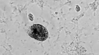
La photo de la semaine : un « Hulk » microscopique filmé en train de se déchaîner contre les siens !
écaille comportement
Et si Hulk existait… à l’échelle microscopique ? Derrière cette image fascinante se cache un comportement aussi spectaculaire qu’inattendu… qui intrigue les scientifiques.
05/07/2026 futura-terre
Des modèles étonnants de Haolonggood Sarcosuchus arrivent chez Everything Dinosaur
écaille Dinosauria
Les modèles très attendus de Haolonggood Sarcosuchus imperator sont arrivés dans l’entrepôt d’Everything Dinosaur. Deux superbes répliques à l'échelle 1/35 de l'énorme crocodile préhistorique Sarcosuchus sont désormais en stock. Les collectionneurs peuvent choisir entre deux variantes de couleurs saisissantes. Le Haolonggood Sarcosuchus imperator Tong Meng présente une belle coloration brune. En revanche, l'empereur Haolonggood Sarcosuchus Tong Wei
03/07/2026 everythingdinosaur ⚙ Traduction automatique
Le nouveau modèle Haolonggood Coelodonta arrive chez Everything Dinosaur
membre écaille Dinosauria mammifères
Le très attendu modèle Haolonggood Coelodonta antiquitatis est arrivé dans l’entrepôt d’Everything Dinosaur. Cette magnifique réplique à l'échelle 1:35 représente l'un des mammifères préhistoriques les plus célèbres de la période glaciaire : l'emblématique rhinocéros laineux. Modèle Haolonggood Woolly Rhino à l'échelle 1:35. Les membres de l'équipe ont été occupés à vérifier le nouvel arrivant et à préparer
02/07/2026 everythingdinosaur ⚙ Traduction automatique
125-Million-Year-Old Crocodile Relative Reveals Its True Colors
Un parent de crocodile âgé de 125 millions d'années révèle ses vraies couleurs
écaille peau Espagne Crétacé Crétacé inférieur autres reptiles
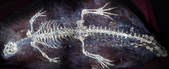
La peau exceptionnellement préservée de Montsecosuchus depereti, un crocodylomorphe éteint pas plus gros qu'un chat domestique qui rôdait dans les zones humides tropicales de l'Espagne du Crétacé inférieur, a permis aux paléontologues de reconstituer les détails de ses écailles, de ses organes sensoriels et même d'éventuelles marques en bandes le long de sa queue. L'article Un parent de crocodile âgé de 125 millions d'années révèle ses vraies couleurs est apparu en premier sur Sci.News : Breaking Science News.
18/06/2026 sci-news ⚙ Traduction automatique

Voici la première carte mondiale d’un immense réseau vivant caché sous nos pieds
écaille étude
Sous nos pieds se cache l’un des plus vastes réseaux vivants de la planète. Une nouvelle étude révèle l’ampleur vertigineuse des champignons mycorhiziens, ces alliés invisibles des plantes qui transportent des milliards de tonnes de carbone chaque année et pourraient jouer un rôle clé dans la...
13/06/2026 futura-terre
1 2 3 4 5