vertèbre

Partie anatomique

80 image(s) · 11 Actualités

Galerie d'images

Cervical vertebra (Cv 12) of Austroposeidon magnificus gen. et nov. sp.

(A) Left lateral and (B) anterior views. Abbreviations: acdl, anterior centrodiapophyseal lamina; acpl, anterior centroparapophyseal lamina; cprl, centroprezygapophyseal lamina; d, diapophysis; prz, prezygapophysis; prdl, prezygodiapophyseal lamina; prsl, prespinal lamina; s, neural spine; sprl, spinoprezygapophyseal lamina. Scale bar: 100mm.
Taxons Austroposeidon

Cervical vertebra (Cv 12) of Austroposeidon magnificus gen. et nov. sp. (A) Left lateral and (B) anterior views. Abbreviations: acdl, anterior centrodiapophyseal lamina; acpl, anterior centroparapophyseal lamina; cprl, centroprezygapophyseal lamina; d, diapophysis; prz, prezygapophysis; prdl, prezygodiapophyseal lamina; prsl, prespinal lamina; s, neural spine; sprl, spinoprezygapophyseal lamina. Scale bar: 100mm.

écaille vertèbre Austroposeidon
Bones referred to Ibirania parva, A. Middle caudal vertebra (MPMA 08-0060-07) referred to I. parva. B. Views of partial right ulna (LPP-PV-0202) of the I. parva holotype. According to Navarro et al. (2022).

Bones referred to Ibirania parva, A. Middle caudal vertebra (MPMA 08-0060-07) referred to I. parva. B. Views of partial right ulna (LPP-PV-0202) of the I. parva holotype. According to Navarro et al. (2022).

os vertèbre holotype Ibirania +1
Representative taxa from the Santonian Iharkút fauna from the Csehbánya Formation, Bakony Mountains, western Hungary. A Pannoniasaurus inexpectatus (Squamata, Mosasauroidea), dorsal vertebra (MTM uncatalogued) in dorsal view (photo by Réka Kalmár) B Foxemys trabanti (Pleurodira, Bothremydidae), skull (MTM V 2010.215.1.) in dorsal view (photo by Márton Rabi). C Bicuspidon aff. hatzegiensis (Squamata, Borioteiioidea), left dentary (MTM 2006.112.1.) in medial view (photo by László Makádi) D Basal tetanuran (Theropoda, Tetanurae), tooth (MTM V.01.54) in ?lingual view E Indeterminate abelisaurid (Theropoda, Abelisauridae), pedal ungual phalanx (MTM V 2008.43.1.) in lateral view F Pneumatoraptor fodori (Theropoda, Paraves), left scapulocoracoid (holotype, MTM V 2008.38.1.) in lateral view G Mochlodon vorosi (Ornithopoda, Rhabdodontidae), left dentary (holotype, MTM V 2010.105.1) in lateral view H Bakonydraco galaczi (Pterosauria, Azhdarchidae), mandible (holotype, MTM 2007.110.1) in dorsal view I Iharkutosuchus makadii (Eusuchia, Hylaeochampsidae), skull (holotype, MTM 2006.52.1) in dorsal view J Hungarosaurus tormai (Ankylosauria, Nodosauridae), right dentary (MTM 2007.25.2) in lateral view K Bauxitornis mindszentyae (Aves, Enantiornithes), left tarsometatarsus (holotype, MTM V 2009.38.1) in anterior view L Ajkaceratops kozmai (Ceratopsia), fused rostral and premaxillae (holotype, MTM V 2009.192.1) in lateral view. Scale bars: 2 cm in A, V, G, H, I, J; 1 cm in D, E, F, K, L; 1 mm in C.

Representative taxa from the Santonian Iharkút fauna from the Csehbánya Formation, Bakony Mountains, western Hungary. A Pannoniasaurus inexpectatus (Squamata, Mosasauroidea), dorsal vertebra (MTM uncatalogued) in dorsal view (photo by Réka Kalmár) B Foxemys trabanti (Pleurodira, Bothremydidae), skull (MTM V 2010.215.1.) in dorsal view (photo by Márton Rabi). C Bicuspidon aff. hatzegiensis (Squamata, Borioteiioidea), left dentary (MTM 2006.112.1.) in medial view (photo by László Makádi) D Basal tetanuran (Theropoda, Tetanurae), tooth (MTM V.01.54) in ?lingual view E Indeterminate abelisaurid (Theropoda, Abelisauridae), pedal ungual phalanx (MTM V 2008.43.1.) in lateral view F Pneumatoraptor fodori (Theropoda, Paraves), left scapulocoracoid (holotype, MTM V 2008.38.1.) in lateral view G Mochlodon vorosi (Ornithopoda, Rhabdodontidae), left dentary (holotype, MTM V 2010.105.1) in lateral view H Bakonydraco galaczi (Pterosauria, Azhdarchidae), mandible (holotype, MTM 2007.110.1) in dorsal view I Iharkutosuchus makadii (Eusuchia, Hylaeochampsidae), skull (holotype, MTM 2006.52.1) in dorsal view J Hungarosaurus tormai (Ankylosauria, Nodosauridae), right dentary (MTM 2007.25.2) in lateral view K Bauxitornis mindszentyae (Aves, Enantiornithes), left tarsometatarsus (holotype, MTM V 2009.38.1) in anterior view L Ajkaceratops kozmai (Ceratopsia), fused rostral and premaxillae (holotype, MTM V 2009.192.1) in lateral view. Scale bars: 2 cm in A, V, G, H, I, J; 1 cm in D, E, F, K, L; 1 mm in C.

dent vertèbre Hongrie Santonien +14
MRF 319, a partial oviraptorosaurian skeleton referred to Anzu wyliei. 
(A) Skeletal reconstruction in left lateral view, with preserved bones in gray and bones represented in other Anzu specimens in white (hatching indicates heavily reconstructed portions of the ilia of CM 78001). Middle-posterior (ninth?) cervical vertebra in (B) anterior, (C) left lateral, and (D) dorsal views. Posterior (11th?) cervical vertebra in (E) anterior, (F) left lateral, and (G) dorsal views. Posterior (12th?) cervical vertebra in (H) anterior, (I) left lateral, and (J) dorsal views. Anteroposteriorly crushed left radius in lateral (K) and anterior (L) views. Mediolaterally crushed left ulna in lateral (M) and anterior (N) views. (O) Partial left scapulocoracoid in lateral view. Dorsal rib in anterior (P) and posterior (Q) views. Abbreviations: acr, acromial process; cr, cervical rib; dip, distal processes; pat, pathology; pf, pneumatic fossa. Scale bars = 50 cm in A; 1 cm in B–Q.

MRF 319, a partial oviraptorosaurian skeleton referred to Anzu wyliei. (A) Skeletal reconstruction in left lateral view, with preserved bones in gray and bones represented in other Anzu specimens in white (hatching indicates heavily reconstructed portions of the ilia of CM 78001). Middle-posterior (ninth?) cervical vertebra in (B) anterior, (C) left lateral, and (D) dorsal views. Posterior (11th?) cervical vertebra in (E) anterior, (F) left lateral, and (G) dorsal views. Posterior (12th?) cervical vertebra in (H) anterior, (I) left lateral, and (J) dorsal views. Anteroposteriorly crushed left radius in lateral (K) and anterior (L) views. Mediolaterally crushed left ulna in lateral (M) and anterior (N) views. (O) Partial left scapulocoracoid in lateral view. Dorsal rib in anterior (P) and posterior (Q) views. Abbreviations: acr, acromial process; cr, cervical rib; dip, distal processes; pat, pathology; pf, pneumatic fossa. Scale bars = 50 cm in A; 1 cm in B–Q.

os écaille vertèbre pathologie +5
Plate XII(XIX).
Fig. 1. Coelophysis bauri COPE. Sacrum, consisting of three vertebrae and last dorsal vertebra. COPE’s original. Triassic, New Mexico. Preserved in American Museum of Natural History, New York. Nat. size, from a cast in Tübingen. a, right lateral view; b, left lateral view; c, ventral view.
Fig. 2. Thecodontosaurus skirtopodus SEELEY sp. Right humerus. Original in Vienna Hofmuseum (Coll. ADLER 1886). Upper Karroo, Cape Colony, South Africa. 1/2 nat. size, from a cast in Tübingen.
Fig. 3. Same. Ditto. Proximal end of a right humerus in posterior view. 1/2 nat. size (the lateral part is missing).
Fig. 4. Same. Ditto. Distal end of a left humerus in anterior view. 1/2 nat. size.
Fig. 5. Same. Ditto. Distal end of a left femur in posterior view. 1/2 nat. size.
Fig. 6. Same. Ditto. Proximal end of a left tibia, lateral view. 1/2 nat. size.
Fig. 7. Thecodontosaurus browni SEELEY sp. Left femur in posterior view. SEELEY’s original. From the Stormberg Beds of the Telle River near Aliwal North, Cape Colony, South Africa. (From casts in the British Museum and Tübingen.) 1/2 nat. size.

Fig. 8. Same. Ditto. Right femur, medial view.

Plate XII(XIX). Fig. 1. Coelophysis bauri COPE. Sacrum, consisting of three vertebrae and last dorsal vertebra. COPE’s original. Triassic, New Mexico. Preserved in American Museum of Natural History, New York. Nat. size, from a cast in Tübingen. a, right lateral view; b, left lateral view; c, ventral view. Fig. 2. Thecodontosaurus skirtopodus SEELEY sp. Right humerus. Original in Vienna Hofmuseum (Coll. ADLER 1886). Upper Karroo, Cape Colony, South Africa. 1/2 nat. size, from a cast in Tübingen. Fig. 3. Same. Ditto. Proximal end of a right humerus in posterior view. 1/2 nat. size (the lateral part is missing). Fig. 4. Same. Ditto. Distal end of a left humerus in anterior view. 1/2 nat. size. Fig. 5. Same. Ditto. Distal end of a left femur in posterior view. 1/2 nat. size. Fig. 6. Same. Ditto. Proximal end of a left tibia, lateral view. 1/2 nat. size. Fig. 7. Thecodontosaurus browni SEELEY sp. Left femur in posterior view. SEELEY’s original. From the Stormberg Beds of the Telle River near Aliwal North, Cape Colony, South Africa. (From casts in the British Museum and Tübingen.) 1/2 nat. size. Fig. 8. Same. Ditto. Right femur, medial view.

humérus vertèbre musée Mexique +8
Complete specimen, excellent mineralization. Presented with a base (exceptional, very good condition)).	
Skull length 18,9 inch ( 28,3 inch with vertebras)

Complete specimen, excellent mineralization. Presented with a base (exceptional, very good condition)). Skull length 18,9 inch ( 28,3 inch with vertebras)

vertèbre spécimen Platecarpus crâne
Dorsal vertebra of platecarpus, a cretaceous. Mosasaur from the Niobrara Chalk of Kansas etc.

General Collections
Keywords: prehistoric archaeology; Paleopathology; Moodie, Roy Lee

Dorsal vertebra of platecarpus, a cretaceous. Mosasaur from the Niobrara Chalk of Kansas etc. General Collections Keywords: prehistoric archaeology; Paleopathology; Moodie, Roy Lee

vertèbre Niobrara Crétacé Platecarpus
Atsinganosaurus velauciensis (VBN 93.01), late Campanian, Velaux-La Bastide Neuve, Bouches-de-Rhône, southern France. Posterior dorsal vertebra, right lateral view. Scale bar equals 10 cm.
Taxons Atsinganosaurus

Atsinganosaurus velauciensis (VBN 93.01), late Campanian, Velaux-La Bastide Neuve, Bouches-de-Rhône, southern France. Posterior dorsal vertebra, right lateral view. Scale bar equals 10 cm.

écaille vertèbre France Campanien +1
Middle cervical vertebra of Vouivria damparisensis (MNHN.F.1934.6 DAM 6).

(A) Left lateral view, (B) anterior view, (C) right lateral view; (D) posterior view. Abbreviations: cpol, centropostzygapophyseal lamina; di, diapophysis; cprl, centroprezygapophyseal lamina; espol, expanded spinopostzygapophyseal lamina; no, notch; ns, notch; pa, parapophysis; pn, pneumatic foramen; pocdf, postzygapophyseal centrodiapophyseal fossa; podl, postzygodiapophyseal lamina; poz, postzygapophysis; prz, prezygapophysis; ri, ridge; sdf, spinodiapophyseal fossa; spol, spinopostzygapophyseal lamina; sprl, spinoprezygapophyseal lamina. Scale bar equals 10 cm.
Taxons Vouivria

Middle cervical vertebra of Vouivria damparisensis (MNHN.F.1934.6 DAM 6). (A) Left lateral view, (B) anterior view, (C) right lateral view; (D) posterior view. Abbreviations: cpol, centropostzygapophyseal lamina; di, diapophysis; cprl, centroprezygapophyseal lamina; espol, expanded spinopostzygapophyseal lamina; no, notch; ns, notch; pa, parapophysis; pn, pneumatic foramen; pocdf, postzygapophyseal centrodiapophyseal fossa; podl, postzygodiapophyseal lamina; poz, postzygapophysis; prz, prezygapophysis; ri, ridge; sdf, spinodiapophyseal fossa; spol, spinopostzygapophyseal lamina; sprl, spinoprezygapophyseal lamina. Scale bar equals 10 cm.

écaille vertèbre Vouivria
Fig. 1. Type material of Amygdalodon patagonicus Cabrera. 1947. A. B. Tooth, MLP 46-VIII-21-1/13, in lingual (A) and labial (B) view. C. D, E. Cervical vertebra, MLP 46-VIII-21-1/8. in right lateral (C, stereopair), ventral (D) and left lateral (E) view. F, G. Right cervical prezygapophysis, MLP 46-VIII-21-117. in dorsal (F) and medial (G) view. H. Anterior dorsal neural spine, MLP 46-VIII-21-116. in posterior view. I, J, K. Posterior dorsal vertebra, MLP 46-VIII-21-112, lectotype, in right lateral view (I, stereopair), right lateral view, with right wall of neural canal remived (J), and posterior (K) view. L, M. Caudal vertebra, MLP 46-VIII-21-1/3, in left lateral (L) and posterior (M) view. N. Caudal vertebra, MLP 46-VIII-21-1/4, in right lateral view. (O). Proximal fragment of a dorsal rib, MLP 46-VIII-21-1/9, in anterolateral view. Abbreviations: acdl, anterior centrodiapophyseal lamina; cprl, centroprezygapophyseal lamina; g, groove; k, ventral keel; pcdl, posterior centrodiapophyseal lamina; pod, postzygodiapophyseal lamina; poz, postzygapophysis; pp, parapophysis; prdl, prezygodiapophyseal lamina; spol, spinopostzgapophyseal lamina; sprl, spinoprezygapophyseal lamina; tprl, intraprezygapophyseal lamina. Scale bars 1 cm (A, B) and 10 cm (C-0).
Taxons Amygdalodon

Fig. 1. Type material of Amygdalodon patagonicus Cabrera. 1947. A. B. Tooth, MLP 46-VIII-21-1/13, in lingual (A) and labial (B) view. C. D, E. Cervical vertebra, MLP 46-VIII-21-1/8. in right lateral (C, stereopair), ventral (D) and left lateral (E) view. F, G. Right cervical prezygapophysis, MLP 46-VIII-21-117. in dorsal (F) and medial (G) view. H. Anterior dorsal neural spine, MLP 46-VIII-21-116. in posterior view. I, J, K. Posterior dorsal vertebra, MLP 46-VIII-21-112, lectotype, in right lateral view (I, stereopair), right lateral view, with right wall of neural canal remived (J), and posterior (K) view. L, M. Caudal vertebra, MLP 46-VIII-21-1/3, in left lateral (L) and posterior (M) view. N. Caudal vertebra, MLP 46-VIII-21-1/4, in right lateral view. (O). Proximal fragment of a dorsal rib, MLP 46-VIII-21-1/9, in anterolateral view. Abbreviations: acdl, anterior centrodiapophyseal lamina; cprl, centroprezygapophyseal lamina; g, groove; k, ventral keel; pcdl, posterior centrodiapophyseal lamina; pod, postzygodiapophyseal lamina; poz, postzygapophysis; pp, parapophysis; prdl, prezygodiapophyseal lamina; spol, spinopostzgapophyseal lamina; sprl, spinoprezygapophyseal lamina; tprl, intraprezygapophyseal lamina. Scale bars 1 cm (A, B) and 10 cm (C-0).

dent vertèbre Amygdalodon
Cervical vertebra of Phosphatodraco mauritanicus (FSAC-OB 12) in (A) dorsal view, (B) ventral view, (C) left lateral view; (D) anterior view; (E) posterior view. Abbreviations: cot = cotyle; hyp = hypapophysis; nsp = neural spine; pex = postexapophysis; poz, postzygapophysis; prz, prezygapophysis.

Cervical vertebra of Phosphatodraco mauritanicus (FSAC-OB 12) in (A) dorsal view, (B) ventral view, (C) left lateral view; (D) anterior view; (E) posterior view. Abbreviations: cot = cotyle; hyp = hypapophysis; nsp = neural spine; pex = postexapophysis; poz, postzygapophysis; prz, prezygapophysis.

vertèbre Phosphatodraco
Eopteranodon lii holotype (BPV 078).

(A) Counterpart; (B) main part. (C and D) Respective schematic drawings. Abbreviations: cv, cervical vertebra; co, coracoid; d1–d4, digits 1–4; fe, femur; fi, fibula; h, humerus; j, jugal; mand, mandible; mc, metacarpal; pmc, premaxillary crest; pe, pelvis; ph, phalanx; ti, tibia; ul, ulna; rad, radius. Scale bars: C, 50 mm; D, 10 mm.

Eopteranodon lii holotype (BPV 078). (A) Counterpart; (B) main part. (C and D) Respective schematic drawings. Abbreviations: cv, cervical vertebra; co, coracoid; d1–d4, digits 1–4; fe, femur; fi, fibula; h, humerus; j, jugal; mand, mandible; mc, metacarpal; pmc, premaxillary crest; pe, pelvis; ph, phalanx; ti, tibia; ul, ulna; rad, radius. Scale bars: C, 50 mm; D, 10 mm.

crête humérus bassin écaille +4
Fig. 11. Laevisuchus indicus (K20/613), cervical vertebra in A, posterior, B, right lateral, C, anterior, and D, dorsal views. Abbreviations: dp, diapophysis; ep, epipophysis; nc, neural canal; ns, neural spine; pf, pneumatic fossa; pl, pleurncoel; poz, postzygapophysis; pp, parapophysis; prz, prezygapophysis.

Fig. 11. Laevisuchus indicus (K20/613), cervical vertebra in A, posterior, B, right lateral, C, anterior, and D, dorsal views. Abbreviations: dp, diapophysis; ep, epipophysis; nc, neural canal; ns, neural spine; pf, pneumatic fossa; pl, pleurncoel; poz, postzygapophysis; pp, parapophysis; prz, prezygapophysis.

vertèbre Laevisuchus
Skeletal diagram of Veterupristisaurus milneri including all known remains. The middle vertebrae is the holotype (MB R 1938), the pair on the right are the paratypes (MB R 2166), and the far left caudal is the questionably referred vertebra (MB R 1940).

Skeletal diagram of Veterupristisaurus milneri including all known remains. The middle vertebrae is the holotype (MB R 1938), the pair on the right are the paratypes (MB R 2166), and the far left caudal is the questionably referred vertebra (MB R 1940).

vertèbre holotype Veterupristisaurus
Cranial and postcranial sauropod remains from sediments of the Praia da Amoreira-Porto Novo Fm. of the coastal sector of Praia da Consolação-Lourinhã-Torres Vedras: 1-2, ?Turiasauria indet., heart-shaped tooth (SHN (JJS) 142, Praia da Corva) in lingual (1) and labial (2) views; 3-4, ?Macronaria indet., spatulate tooth (SHN 513, Porto Novo) in labial (3) and lingual (4) views; 5-6, Macronaria indet., compressed cone-chisel-shaped tooth (SHN 578, Valmitão) in lingual (5) and labial (6) views; 7-8, Eusauropoda indet., partial left maxilla (SHN 582, Praia dos Frades) in lateral (7) and posterior (8) views; 9, Titanosauriformes indet., posterior caudal vertebra (SHN 523, Praia da Corva) in right view; 10-12, Diplodocinae indet., partial skeleton (SHN (JJS) 177, Valmitão), anterior caudal neural spine in posterior view (10), anterior caudal centrum in right view (11) and left ischium in medial view (12); 13-16, holotype material of Zby atlanticus (ML 368, Vale de Pombas), right ungueal I in lateral view (13), right humerus in anterior view (14), right radius in posterior view (15), right ulna in lateral view (16); 17, Eusauropoda indet., partial distal forked-chevron (SHN 587, Praia da Corva) in medial view; 18, Sauropoda indet., pedal ungueal I (SHN 524, Praia de Pedrogãos) in lateral view; 19-22, Macronarian indet., partial skeleton (SHN 181, Valmitão), right astragalus in proximal view (19), anterior caudal vertebra in anterior view (20), right tibia in lateral view (21) and right fibula in medial view (22); 23-24, Eusauropoda indet., partial skeleton (SHN 530, Praia da Corva), anterior chevron in anterior view (23) and anterior caudal vertebra in posterior view (24); 25, cf. Duriatitan humerocristatus, humerus (MG 4976, Praia dos Frades) in anterior view; 26, Sauropoda indet., partial skeleton (SHN 534, Santa Rita), middle chevron in posterior view; 27, Diplodocidae indet., partial skeleton (SHN (JJS) 179, Praia Vermelha), dorsal/caudal (?) neural spine in posterior view; 28-30, holotype material of Dinheirosaurus lourinhanensis (ML 414, Porto Dinheiro), proximal end of a dorsal rib in anterior view (28), anterior caudal neural spine in posterior view (29) and articulated dorsal vertebrae in right view (30). Black scale bar: 10 cm; Grey scale bar: 5 cm; Brown scale bar: 1 cm. See Anatomical abbreviations for abbreviations.

Cranial and postcranial sauropod remains from sediments of the Praia da Amoreira-Porto Novo Fm. of the coastal sector of Praia da Consolação-Lourinhã-Torres Vedras: 1-2, ?Turiasauria indet., heart-shaped tooth (SHN (JJS) 142, Praia da Corva) in lingual (1) and labial (2) views; 3-4, ?Macronaria indet., spatulate tooth (SHN 513, Porto Novo) in labial (3) and lingual (4) views; 5-6, Macronaria indet., compressed cone-chisel-shaped tooth (SHN 578, Valmitão) in lingual (5) and labial (6) views; 7-8, Eusauropoda indet., partial left maxilla (SHN 582, Praia dos Frades) in lateral (7) and posterior (8) views; 9, Titanosauriformes indet., posterior caudal vertebra (SHN 523, Praia da Corva) in right view; 10-12, Diplodocinae indet., partial skeleton (SHN (JJS) 177, Valmitão), anterior caudal neural spine in posterior view (10), anterior caudal centrum in right view (11) and left ischium in medial view (12); 13-16, holotype material of Zby atlanticus (ML 368, Vale de Pombas), right ungueal I in lateral view (13), right humerus in anterior view (14), right radius in posterior view (15), right ulna in lateral view (16); 17, Eusauropoda indet., partial distal forked-chevron (SHN 587, Praia da Corva) in medial view; 18, Sauropoda indet., pedal ungueal I (SHN 524, Praia de Pedrogãos) in lateral view; 19-22, Macronarian indet., partial skeleton (SHN 181, Valmitão), right astragalus in proximal view (19), anterior caudal vertebra in anterior view (20), right tibia in lateral view (21) and right fibula in medial view (22); 23-24, Eusauropoda indet., partial skeleton (SHN 530, Praia da Corva), anterior chevron in anterior view (23) and anterior caudal vertebra in posterior view (24); 25, cf. Duriatitan humerocristatus, humerus (MG 4976, Praia dos Frades) in anterior view; 26, Sauropoda indet., partial skeleton (SHN 534, Santa Rita), middle chevron in posterior view; 27, Diplodocidae indet., partial skeleton (SHN (JJS) 179, Praia Vermelha), dorsal/caudal (?) neural spine in posterior view; 28-30, holotype material of Dinheirosaurus lourinhanensis (ML 414, Porto Dinheiro), proximal end of a dorsal rib in anterior view (28), anterior caudal neural spine in posterior view (29) and articulated dorsal vertebrae in right view (30). Black scale bar: 10 cm; Grey scale bar: 5 cm; Brown scale bar: 1 cm. See Anatomical abbreviations for abbreviations.

dent vertèbre Lourinhã holotype +7
Anterior dorsal vertebra of the sauropod dinosaur Tendaguria tanzaniensis.
Taxons Tendaguriidae

Anterior dorsal vertebra of the sauropod dinosaur Tendaguria tanzaniensis.

vertèbre Dinosauria Tendaguria Tendaguriidae
1 2 3 4 5

Actualités

Des scientifiques redécouvrent des fossiles perdus de Megalodon et révèlent un géant de 79 pieds
vertèbre musée fossile
Plusieurs énormes vertèbres de Megalodon, qui auraient été détruites en 1989, ont été retrouvées inaperçues sur une étagère de musée. Leur analyse renforce la preuve que le requin géant pourrait dépasser 24 mètres (79 pieds) de longueur et vivre près d'un siècle. Les chercheurs estiment également que ses nouveau-nés auraient pu mesurer une taille étonnante de 3,6 mètres (12 pieds).
07/08/2026 sciencedaily ⚙ Traduction automatique
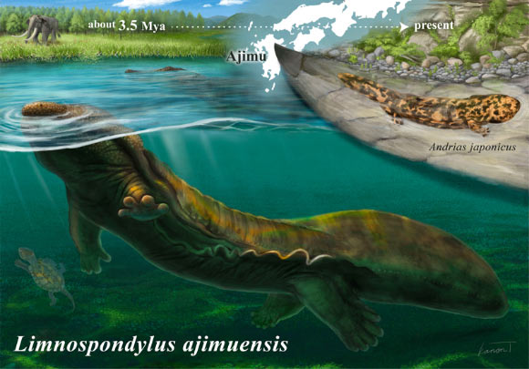
3.5-Million-Year-Old Fossil from Japan Reveals Lost Lineage of Giant Salamanders
Un fossile vieux de 3,5 millions d'années provenant du Japon révèle la lignée perdue de salamandres géantes
vertèbre Japon fossile nouvelle espèce
Un nouveau genre et une nouvelle espèce de salamandre cryptobranchidée qui vivait dans les lacs du Japon il y a environ 3,5 millions d’années ont été identifiés à partir de trois vertèbres fossiles, comblant ainsi une lacune majeure dans les archives fossiles de l’une des plus anciennes lignées d’amphibiens au monde. L'article Un fossile vieux de 3,5 millions d'années en provenance du Japon révèle une lignée perdue de salamandres géantes est apparu en premier sur Sci.News : Breaking Science News.
21/07/2026 sci-news ⚙ Traduction automatique
Uragasaurus – Un nouveau sauropode Mamenchisaurid de Thaïlande
os membre vertèbre Thaïlande fossile Dinosauria Mamenchisauridae Uragasaurus nouvelle espèce
Une équipe de chercheurs internationaux a nommé une nouvelle espèce remarquable de dinosaure à long cou originaire de Thaïlande. Uragasaurus kalasinensis est le premier membre formellement décrit des Mamenchisauridae de Thaïlande. Bien que les preuves fossiles consistent en une seule vertèbre, l'os préserve une combinaison unique de caractéristiques anatomiques qui ont permis aux scientifiques d'identifier un tout nouveau corps.
15/07/2026 everythingdinosaur ⚙ Traduction automatique
83-Million-Year-Old Fossil Rewrites Timeline of Antarctica’s First Dinosaur Discovery
Un fossile vieux de 83 millions d’années réécrit la chronologie de la première découverte de dinosaures en Antarctique
vertèbre Antarctique fossile Dinosauria Titanosauria découverte
Un petit fossile collecté sur une île de l'Antarctique il y a plus de quatre décennies est la vertèbre caudale d'un dinosaure sauropode titanosaure qui parcourait l'Antarctique il y a environ 83 millions d'années, selon un nouvel article paru dans la revue Acta Palaeontologica Polonica. La chronologie de la première découverte de dinosaures en Antarctique réécrite par un fossile vieux de 83 millions d'années est apparue en premier sur Sci.News : Breaking Science News.
01/07/2026 sci-news ⚙ Traduction automatique
Cet os oublié dans un tiroir depuis 40 ans vient de révéler le tout premier dinosaure découvert en Antarctique !
os vertèbre Antarctique Crétacé supérieur fossile Dinosauria Titanosauria découverte
Pendant près de quarante ans, il est resté oublié dans un tiroir du British Antarctic Survey. Ce fossile, découvert en Antarctique en 1985, vient pourtant d'être identifié comme le tout premier fossile de dinosaure jamais mis au jour sur le continent blanc. Une découverte qui éclaire aussi les...
01/07/2026 futura-terre
1 2 3