vertebra

Anatomical part

80 image(s) · 11 News

Image gallery

Cervical vertebra of Phosphatodraco mauritanicus (FSAC-OB 12) in (A) dorsal view, (B) ventral view, (C) left lateral view; (D) anterior view; (E) posterior view. Abbreviations: cot = cotyle; hyp = hypapophysis; nsp = neural spine; pex = postexapophysis; poz, postzygapophysis; prz, prezygapophysis.
Taxa Phosphatodraco

Cervical vertebra of Phosphatodraco mauritanicus (FSAC-OB 12) in (A) dorsal view, (B) ventral view, (C) left lateral view; (D) anterior view; (E) posterior view. Abbreviations: cot = cotyle; hyp = hypapophysis; nsp = neural spine; pex = postexapophysis; poz, postzygapophysis; prz, prezygapophysis.

vertebra Phosphatodraco
Eopteranodon lii holotype (BPV 078).

(A) Counterpart; (B) main part. (C and D) Respective schematic drawings. Abbreviations: cv, cervical vertebra; co, coracoid; d1–d4, digits 1–4; fe, femur; fi, fibula; h, humerus; j, jugal; mand, mandible; mc, metacarpal; pmc, premaxillary crest; pe, pelvis; ph, phalanx; ti, tibia; ul, ulna; rad, radius. Scale bars: C, 50 mm; D, 10 mm.
Taxa Eopteranodon

Eopteranodon lii holotype (BPV 078). (A) Counterpart; (B) main part. (C and D) Respective schematic drawings. Abbreviations: cv, cervical vertebra; co, coracoid; d1–d4, digits 1–4; fe, femur; fi, fibula; h, humerus; j, jugal; mand, mandible; mc, metacarpal; pmc, premaxillary crest; pe, pelvis; ph, phalanx; ti, tibia; ul, ulna; rad, radius. Scale bars: C, 50 mm; D, 10 mm.

crest humerus pelvis scale +4
Cranial and postcranial sauropod remains from sediments of the Praia da Amoreira-Porto Novo Fm. of the coastal sector of Praia da Consolação-Lourinhã-Torres Vedras: 1-2, ?Turiasauria indet., heart-shaped tooth (SHN (JJS) 142, Praia da Corva) in lingual (1) and labial (2) views; 3-4, ?Macronaria indet., spatulate tooth (SHN 513, Porto Novo) in labial (3) and lingual (4) views; 5-6, Macronaria indet., compressed cone-chisel-shaped tooth (SHN 578, Valmitão) in lingual (5) and labial (6) views; 7-8, Eusauropoda indet., partial left maxilla (SHN 582, Praia dos Frades) in lateral (7) and posterior (8) views; 9, Titanosauriformes indet., posterior caudal vertebra (SHN 523, Praia da Corva) in right view; 10-12, Diplodocinae indet., partial skeleton (SHN (JJS) 177, Valmitão), anterior caudal neural spine in posterior view (10), anterior caudal centrum in right view (11) and left ischium in medial view (12); 13-16, holotype material of Zby atlanticus (ML 368, Vale de Pombas), right ungueal I in lateral view (13), right humerus in anterior view (14), right radius in posterior view (15), right ulna in lateral view (16); 17, Eusauropoda indet., partial distal forked-chevron (SHN 587, Praia da Corva) in medial view; 18, Sauropoda indet., pedal ungueal I (SHN 524, Praia de Pedrogãos) in lateral view; 19-22, Macronarian indet., partial skeleton (SHN 181, Valmitão), right astragalus in proximal view (19), anterior caudal vertebra in anterior view (20), right tibia in lateral view (21) and right fibula in medial view (22); 23-24, Eusauropoda indet., partial skeleton (SHN 530, Praia da Corva), anterior chevron in anterior view (23) and anterior caudal vertebra in posterior view (24); 25, cf. Duriatitan humerocristatus, humerus (MG 4976, Praia dos Frades) in anterior view; 26, Sauropoda indet., partial skeleton (SHN 534, Santa Rita), middle chevron in posterior view; 27, Diplodocidae indet., partial skeleton (SHN (JJS) 179, Praia Vermelha), dorsal/caudal (?) neural spine in posterior view; 28-30, holotype material of Dinheirosaurus lourinhanensis (ML 414, Porto Dinheiro), proximal end of a dorsal rib in anterior view (28), anterior caudal neural spine in posterior view (29) and articulated dorsal vertebrae in right view (30). Black scale bar: 10 cm; Grey scale bar: 5 cm; Brown scale bar: 1 cm. See Anatomical abbreviations for abbreviations.
Taxa Zby

Cranial and postcranial sauropod remains from sediments of the Praia da Amoreira-Porto Novo Fm. of the coastal sector of Praia da Consolação-Lourinhã-Torres Vedras: 1-2, ?Turiasauria indet., heart-shaped tooth (SHN (JJS) 142, Praia da Corva) in lingual (1) and labial (2) views; 3-4, ?Macronaria indet., spatulate tooth (SHN 513, Porto Novo) in labial (3) and lingual (4) views; 5-6, Macronaria indet., compressed cone-chisel-shaped tooth (SHN 578, Valmitão) in lingual (5) and labial (6) views; 7-8, Eusauropoda indet., partial left maxilla (SHN 582, Praia dos Frades) in lateral (7) and posterior (8) views; 9, Titanosauriformes indet., posterior caudal vertebra (SHN 523, Praia da Corva) in right view; 10-12, Diplodocinae indet., partial skeleton (SHN (JJS) 177, Valmitão), anterior caudal neural spine in posterior view (10), anterior caudal centrum in right view (11) and left ischium in medial view (12); 13-16, holotype material of Zby atlanticus (ML 368, Vale de Pombas), right ungueal I in lateral view (13), right humerus in anterior view (14), right radius in posterior view (15), right ulna in lateral view (16); 17, Eusauropoda indet., partial distal forked-chevron (SHN 587, Praia da Corva) in medial view; 18, Sauropoda indet., pedal ungueal I (SHN 524, Praia de Pedrogãos) in lateral view; 19-22, Macronarian indet., partial skeleton (SHN 181, Valmitão), right astragalus in proximal view (19), anterior caudal vertebra in anterior view (20), right tibia in lateral view (21) and right fibula in medial view (22); 23-24, Eusauropoda indet., partial skeleton (SHN 530, Praia da Corva), anterior chevron in anterior view (23) and anterior caudal vertebra in posterior view (24); 25, cf. Duriatitan humerocristatus, humerus (MG 4976, Praia dos Frades) in anterior view; 26, Sauropoda indet., partial skeleton (SHN 534, Santa Rita), middle chevron in posterior view; 27, Diplodocidae indet., partial skeleton (SHN (JJS) 179, Praia Vermelha), dorsal/caudal (?) neural spine in posterior view; 28-30, holotype material of Dinheirosaurus lourinhanensis (ML 414, Porto Dinheiro), proximal end of a dorsal rib in anterior view (28), anterior caudal neural spine in posterior view (29) and articulated dorsal vertebrae in right view (30). Black scale bar: 10 cm; Grey scale bar: 5 cm; Brown scale bar: 1 cm. See Anatomical abbreviations for abbreviations.

humerus scale tooth vertebra +13
Anterior dorsal vertebra of the sauropod dinosaur Tendaguria tanzaniensis.
Taxa Tendaguria

Anterior dorsal vertebra of the sauropod dinosaur Tendaguria tanzaniensis.

vertebra Dinosauria Tendaguria
Fig. 11. Laevisuchus indicus (K20/613), cervical vertebra in A, posterior, B, right lateral, C, anterior, and D, dorsal views. Abbreviations: dp, diapophysis; ep, epipophysis; nc, neural canal; ns, neural spine; pf, pneumatic fossa; pl, pleurncoel; poz, postzygapophysis; pp, parapophysis; prz, prezygapophysis.
Taxa Laevisuchus

Fig. 11. Laevisuchus indicus (K20/613), cervical vertebra in A, posterior, B, right lateral, C, anterior, and D, dorsal views. Abbreviations: dp, diapophysis; ep, epipophysis; nc, neural canal; ns, neural spine; pf, pneumatic fossa; pl, pleurncoel; poz, postzygapophysis; pp, parapophysis; prz, prezygapophysis.

vertebra Laevisuchus
Skeletal diagram of Veterupristisaurus milneri including all known remains. The middle vertebrae is the holotype (MB R 1938), the pair on the right are the paratypes (MB R 2166), and the far left caudal is the questionably referred vertebra (MB R 1940).
Taxa Veterupristisaurus

Skeletal diagram of Veterupristisaurus milneri including all known remains. The middle vertebrae is the holotype (MB R 1938), the pair on the right are the paratypes (MB R 2166), and the far left caudal is the questionably referred vertebra (MB R 1940).

vertebra holotype Veterupristisaurus
Entire skeleton of Jidapterus edentus (RCPS-030366CY) preserved in a slab mostly in right lateral view.

Abbreviations: aa, the atlas-axis complex; bo, basioccipital; br, braincase; cve, cervical vertebrae; cv3, the third cervical vertebra; cv8, the eighth cervical vertebra; ds, dentaries; dv, dorsal vertebra; dve, dorsal vertebrae; ep, epiphysis; j, jugal; lc, left carpus; lf, left femur; li, left ilium; lis, left ischium; lm, left manual digits; lp, left pubis; lpe, left pes; lpt, left pteroid; lr, left radius; lsc, left scapula-coracoid; lt, left tibia; lta, left tarsus; lu, left ulna; lwmc, left wing metacarpal; lwp1–4; left wing phalanges 1–4; p, pterygoid; pp, postacetabular process of the right ilium; q, quadrate; rc, right carpus; rh, right humerus; rf, right femur; rfi, right fibula; ribs, ribs; ris, right ischium; rm, right manual digits; rmcI, right metacarpal I; rp, right pubis; rpe, right pes; rpt, right pteroid; rr, right radius; rsc, right scapula-coracoid; rt, right tibia; ru, right ulna; rwmc, right wing metacarpal; rwp1–4, right wing phalanges 1–4; rostrum, rostrum; st, sternum; sv, sacral vertebra; sve, sacral vertebrae. Gray shaded areas bound by solid lines indicate preserved bones, dashed lines indicate bones with margins discerned by impression, and lack of lines indicate missing bone or displacement along cracks in the slab.
Taxa Chaoyangopteridae

Entire skeleton of Jidapterus edentus (RCPS-030366CY) preserved in a slab mostly in right lateral view. Abbreviations: aa, the atlas-axis complex; bo, basioccipital; br, braincase; cve, cervical vertebrae; cv3, the third cervical vertebra; cv8, the eighth cervical vertebra; ds, dentaries; dv, dorsal vertebra; dve, dorsal vertebrae; ep, epiphysis; j, jugal; lc, left carpus; lf, left femur; li, left ilium; lis, left ischium; lm, left manual digits; lp, left pubis; lpe, left pes; lpt, left pteroid; lr, left radius; lsc, left scapula-coracoid; lt, left tibia; lta, left tarsus; lu, left ulna; lwmc, left wing metacarpal; lwp1–4; left wing phalanges 1–4; p, pterygoid; pp, postacetabular process of the right ilium; q, quadrate; rc, right carpus; rh, right humerus; rf, right femur; rfi, right fibula; ribs, ribs; ris, right ischium; rm, right manual digits; rmcI, right metacarpal I; rp, right pubis; rpe, right pes; rpt, right pteroid; rr, right radius; rsc, right scapula-coracoid; rt, right tibia; ru, right ulna; rwmc, right wing metacarpal; rwp1–4, right wing phalanges 1–4; rostrum, rostrum; st, sternum; sv, sacral vertebra; sve, sacral vertebrae. Gray shaded areas bound by solid lines indicate preserved bones, dashed lines indicate bones with margins discerned by impression, and lack of lines indicate missing bone or displacement along cracks in the slab.

bone humerus vertebra Chaoyangopteridae +2
The holotype anterior dorsal vertebra of Uragasaurus kalasinensis (PRC 460) in anterior (a) and posterior (b) views. Digital rendering of the specimen in anterior (c), posterior (d), right lateral (e), left lateral (f), dorsal (g), and ventral (h) views. Asterisk refers to an autapomorphic character. The blue highlight indicates the pneumatic fossa and pleurocoel.
Taxa Uragasaurus

The holotype anterior dorsal vertebra of Uragasaurus kalasinensis (PRC 460) in anterior (a) and posterior (b) views. Digital rendering of the specimen in anterior (c), posterior (d), right lateral (e), left lateral (f), dorsal (g), and ventral (h) views. Asterisk refers to an autapomorphic character. The blue highlight indicates the pneumatic fossa and pleurocoel.

vertebra holotype specimen Uragasaurus
The holotype anterior dorsal vertebra of Uragasaurus kalasinensis (PRC 460) in anterior (a) and posterior (b) views. Digital rendering of the specimen in anterior (c), posterior (d), right lateral (e), left lateral (f), dorsal (g), and ventral (h) views. Asterisk refers to an autapomorphic character. The blue highlight indicates the pneumatic fossa and pleurocoel.

The holotype anterior dorsal vertebra of Uragasaurus kalasinensis (PRC 460) in anterior (a) and posterior (b) views. Digital rendering of the specimen in anterior (c), posterior (d), right lateral (e), left lateral (f), dorsal (g), and ventral (h) views. Asterisk refers to an autapomorphic character. The blue highlight indicates the pneumatic fossa and pleurocoel.

vertebra holotype specimen Uragasaurus
Preserved elements of Tataouinea hannibalis (ONM DT 1–48).
A, quarry map showing the orientation of collected elements. B, field photograph of the elements collected at the end of 2011, and C, of elements collected in 2013. Ca, caudal vertebra 1–17; S, sacral vertebra 1–5; r il, right ilium; l il, left ilium.
Taxa Tataouinea

Preserved elements of Tataouinea hannibalis (ONM DT 1–48). A, quarry map showing the orientation of collected elements. B, field photograph of the elements collected at the end of 2011, and C, of elements collected in 2013. Ca, caudal vertebra 1–17; S, sacral vertebra 1–5; r il, right ilium; l il, left ilium.

vertebra Tataouinea
Skeletal reconstruction of Maraapunisaurus fragillimus, with restored dorsal vertebra in posterior view. Scaled from Lavocatisaurus, Agustinia, Comahuesaurus, Amazonsaurus, & other basal rebbachisaurids, with neck extended allometrically following Stevens & Parrish (2006). Human silhouette is myself.
Taxa Comahuesaurus

Skeletal reconstruction of Maraapunisaurus fragillimus, with restored dorsal vertebra in posterior view. Scaled from Lavocatisaurus, Agustinia, Comahuesaurus, Amazonsaurus, & other basal rebbachisaurids, with neck extended allometrically following Stevens & Parrish (2006). Human silhouette is myself.

vertebra Agustinia Amazonsaurus Comahuesaurus +3
Istiorachis macarthurae holotype (MIWG 6643). The anterior-most seven caudal vertebrae in left lateral view with neural spines reconstructed. Abbreviations: Cd, caudal vertebra; SC, sacrocaudal. Scale bar represents 50 mm.
Taxa Istiorachis

Istiorachis macarthurae holotype (MIWG 6643). The anterior-most seven caudal vertebrae in left lateral view with neural spines reconstructed. Abbreviations: Cd, caudal vertebra; SC, sacrocaudal. Scale bar represents 50 mm.

scale vertebra holotype Istiorachis
Identifier: annalsofmedicalh01pack (find matches)
Title: Annals of medical history
Year: 1917 (1910s)
Authors:  Packard, Francis R. (Francis Randolph), 1870-1950
Subjects:  Medicine
Publisher:  New York P.B. Hoeber
Contributing Library:  Gerstein - University of Toronto
Digitizing Sponsor:  University of Toronto

View Book Page: Book Viewer
About This Book: Catalog Entry
View All Images: All Images From Book
Click here to view book online to see this illustration in context in a browseable online version of this book.

Text Appearing Before Image:
ail strongly Hexed, and the toescontracted and appressed. The whole atti-tude strongly suggests a spastic distress,possibly brought on by some form ofpoisoning of the central nervous system,from infection or the deglutition of somepoisonous substance. 14. Osteomalacia is evidently the causeof the hypertrophy of the bones of Lim-nocyron potens, an early carnivore from theWashakie Eocene of Wyoming, nearly3,000,000 years old. MATERIALS AND METHODS The material described in the presentpaper has been loaned the writer lor de-scription by the Field Museum of Chii Osborn: Hull. Amcr. \lus. Natl. Hist., 1917,vol. 35. !>• 733. P>- 28. Studies in Paleopathology 393 by the American Museum of Natural His-tory of New York City, by Walker Museumof the University of Chicago, and by the Uni-versity of Kansas Natural History Museum. made by the well-known petrographic meth-ods so common in all geological labora-tories. The diagnoses, where they are at-tempted, arc made from comparisons of the
Text Appearing After Image:
Fig. 20. The skeleton of Strutbiomimus altus, a small dinosaur from the BeliyRiver series (Cretaceous), Red Deer River, Alberta, Canada, now regarded as ofapproximately the same age as the Judith River series. The unique feature ofthe skull is the total absence of teeth, with a size of skull one-third larger thanthe ostrich and a length of body of about fifteen feet. The position of the skeletonis decidedly that of the opisthotonos which may be regarded as an indication ofdisease. (After Osborn). A beautiful specimen of an osteoma, theonly one known so far, on the vertebra of aKansas Cretaceous mosasaur, was given thewriter by Dr. J. M. Armstrong of St. Paul.The writer expresses his obligations to thegentlemen connected with the above-men-tioned institutions and to Dr. Armstrong.The methods used are a combination ofprocedures in the various lines involved.Microscopic sections, which can be madethin enough for immersion lens study, are material with similar lesions in recent hu-man mat

Note About Images

Please note that these images are extracted from scanned page images that may have been digitally enhanced for readability - coloration and appearance of these illustrations may not perfectly resemble the original work.
Taxa Hexing

Identifier: annalsofmedicalh01pack (find matches) Title: Annals of medical history Year: 1917 (1910s) Authors: Packard, Francis R. (Francis Randolph), 1870-1950 Subjects: Medicine Publisher: New York P.B. Hoeber Contributing Library: Gerstein - University of Toronto Digitizing Sponsor: University of Toronto View Book Page: Book Viewer About This Book: Catalog Entry View All Images: All Images From Book Click here to view book online to see this illustration in context in a browseable online version of this book. Text Appearing Before Image: ail strongly Hexed, and the toescontracted and appressed. The whole atti-tude strongly suggests a spastic distress,possibly brought on by some form ofpoisoning of the central nervous system,from infection or the deglutition of somepoisonous substance. 14. Osteomalacia is evidently the causeof the hypertrophy of the bones of Lim-nocyron potens, an early carnivore from theWashakie Eocene of Wyoming, nearly3,000,000 years old. MATERIALS AND METHODS The material described in the presentpaper has been loaned the writer lor de-scription by the Field Museum of Chii Osborn: Hull. Amcr. \lus. Natl. Hist., 1917,vol. 35. !>• 733. P>- 28. Studies in Paleopathology 393 by the American Museum of Natural His-tory of New York City, by Walker Museumof the University of Chicago, and by the Uni-versity of Kansas Natural History Museum. made by the well-known petrographic meth-ods so common in all geological labora-tories. The diagnoses, where they are at-tempted, arc made from comparisons of the Text Appearing After Image: Fig. 20. The skeleton of Strutbiomimus altus, a small dinosaur from the BeliyRiver series (Cretaceous), Red Deer River, Alberta, Canada, now regarded as ofapproximately the same age as the Judith River series. The unique feature ofthe skull is the total absence of teeth, with a size of skull one-third larger thanthe ostrich and a length of body of about fifteen feet. The position of the skeletonis decidedly that of the opisthotonos which may be regarded as an indication ofdisease. (After Osborn). A beautiful specimen of an osteoma, theonly one known so far, on the vertebra of aKansas Cretaceous mosasaur, was given thewriter by Dr. J. M. Armstrong of St. Paul.The writer expresses his obligations to thegentlemen connected with the above-men-tioned institutions and to Dr. Armstrong.The methods used are a combination ofprocedures in the various lines involved.Microscopic sections, which can be madethin enough for immersion lens study, are material with similar lesions in recent hu-man mat Note About Images Please note that these images are extracted from scanned page images that may have been digitally enhanced for readability - coloration and appearance of these illustrations may not perfectly resemble the original work.

bone vertebra museum Canada +10
a Ankylosaurus vertebra from the green sand of Folkestone (UK) - Albian

a Ankylosaurus vertebra from the green sand of Folkestone (UK) - Albian

vertebra United Kingdom Albian Ankylosauria
Representative taxa from the Santonian Iharkút fauna from the Csehbánya Formation, Bakony Mountains, western Hungary. A Pannoniasaurus inexpectatus (Squamata, Mosasauroidea), dorsal vertebra (MTM uncatalogued) in dorsal view (photo by Réka Kalmár) B Foxemys trabanti (Pleurodira, Bothremydidae), skull (MTM V 2010.215.1.) in dorsal view (photo by Márton Rabi). C Bicuspidon aff. hatzegiensis (Squamata, Borioteiioidea), left dentary (MTM 2006.112.1.) in medial view (photo by László Makádi) D Basal tetanuran (Theropoda, Tetanurae), tooth (MTM V.01.54) in ?lingual view E Indeterminate abelisaurid (Theropoda, Abelisauridae), pedal ungual phalanx (MTM V 2008.43.1.) in lateral view F Pneumatoraptor fodori (Theropoda, Paraves), left scapulocoracoid (holotype, MTM V 2008.38.1.) in lateral view G Mochlodon vorosi (Ornithopoda, Rhabdodontidae), left dentary (holotype, MTM V 2010.105.1) in lateral view H Bakonydraco galaczi (Pterosauria, Azhdarchidae), mandible (holotype, MTM 2007.110.1) in dorsal view I Iharkutosuchus makadii (Eusuchia, Hylaeochampsidae), skull (holotype, MTM 2006.52.1) in dorsal view J Hungarosaurus tormai (Ankylosauria, Nodosauridae), right dentary (MTM 2007.25.2) in lateral view K Bauxitornis mindszentyae (Aves, Enantiornithes), left tarsometatarsus (holotype, MTM V 2009.38.1) in anterior view L Ajkaceratops kozmai (Ceratopsia), fused rostral and premaxillae (holotype, MTM V 2009.192.1) in lateral view. Scale bars: 2 cm in A, V, G, H, I, J; 1 cm in D, E, F, K, L; 1 mm in C.

Representative taxa from the Santonian Iharkút fauna from the Csehbánya Formation, Bakony Mountains, western Hungary. A Pannoniasaurus inexpectatus (Squamata, Mosasauroidea), dorsal vertebra (MTM uncatalogued) in dorsal view (photo by Réka Kalmár) B Foxemys trabanti (Pleurodira, Bothremydidae), skull (MTM V 2010.215.1.) in dorsal view (photo by Márton Rabi). C Bicuspidon aff. hatzegiensis (Squamata, Borioteiioidea), left dentary (MTM 2006.112.1.) in medial view (photo by László Makádi) D Basal tetanuran (Theropoda, Tetanurae), tooth (MTM V.01.54) in ?lingual view E Indeterminate abelisaurid (Theropoda, Abelisauridae), pedal ungual phalanx (MTM V 2008.43.1.) in lateral view F Pneumatoraptor fodori (Theropoda, Paraves), left scapulocoracoid (holotype, MTM V 2008.38.1.) in lateral view G Mochlodon vorosi (Ornithopoda, Rhabdodontidae), left dentary (holotype, MTM V 2010.105.1) in lateral view H Bakonydraco galaczi (Pterosauria, Azhdarchidae), mandible (holotype, MTM 2007.110.1) in dorsal view I Iharkutosuchus makadii (Eusuchia, Hylaeochampsidae), skull (holotype, MTM 2006.52.1) in dorsal view J Hungarosaurus tormai (Ankylosauria, Nodosauridae), right dentary (MTM 2007.25.2) in lateral view K Bauxitornis mindszentyae (Aves, Enantiornithes), left tarsometatarsus (holotype, MTM V 2009.38.1) in anterior view L Ajkaceratops kozmai (Ceratopsia), fused rostral and premaxillae (holotype, MTM V 2009.192.1) in lateral view. Scale bars: 2 cm in A, V, G, H, I, J; 1 cm in D, E, F, K, L; 1 mm in C.

tooth vertebra Hungary Santonian +15
Dorsal vertebra of platecarpus, a cretaceous. Mosasaur from the Niobrara Chalk of Kansas etc.

General Collections
Keywords: prehistoric archaeology; Paleopathology; Moodie, Roy Lee
Taxa Platecarpus

Dorsal vertebra of platecarpus, a cretaceous. Mosasaur from the Niobrara Chalk of Kansas etc. General Collections Keywords: prehistoric archaeology; Paleopathology; Moodie, Roy Lee

vertebra Niobrara Cretaceous Platecarpus
1 2 3 4 5

News

Scientists rediscover lost Megalodon fossils—and reveal a 79-foot giant
vertebra museum fossil
Several enormous Megalodon vertebrae thought to have been destroyed in 1989 were found sitting unnoticed on a museum shelf. Their analysis strengthens evidence that the giant shark could exceed 24 meters (79 feet) in length and live for nearly a century. Researchers also estimate that its newborns may have measured an astonishing 3.6 meters (12 feet).
07/08/2026 sciencedaily
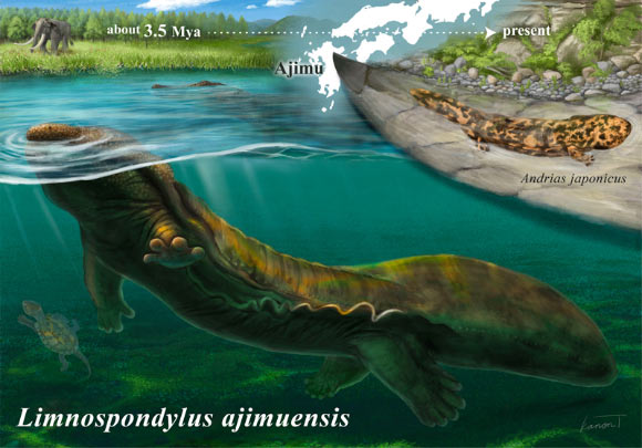
3.5-Million-Year-Old Fossil from Japan Reveals Lost Lineage of Giant Salamanders
vertebra Japan fossil new species
A new genus and species of cryptobranchid salamander that lived in lakes in Japan about 3.5 million years ago has been identified from three fossil vertebrae, filling a major gap in the fossil record of one of the world’s oldest amphibian lineages. The post 3.5-Million-Year-Old Fossil from Japan Reveals Lost Lineage of Giant Salamanders appeared first on Sci.News: Breaking Science News.
21/07/2026 sci-news
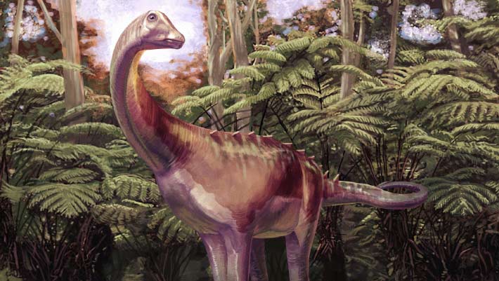
Uragasaurus – A New Mamenchisaurid Sauropod from Thailand
bone limb vertebra Thailand fossil Dinosauria Mamenchisauridae Uragasaurus new species
A team of international researchers has named a remarkable new species of long-necked dinosaur from Thailand. Uragasaurus kalasinensis is the first formally described member of the Mamenchisauridae from Thailand. Although the fossil evidence consists of a single vertebra, the bone preserves a unique combination of anatomical features that enabled scientists to identify an entirely new
15/07/2026 everythingdinosaur
83-Million-Year-Old Fossil Rewrites Timeline of Antarctica’s First Dinosaur Discovery
vertebra Antarctica fossil Dinosauria Titanosauria discovery
A small fossil collected on an Antarctic island more than four decades ago is a tail vertebra of a titanosaurian sauropod dinosaur that roamed Antarctica roughly 83 million years ago, according to a new paper in the journal Acta Palaeontologica Polonica. The post 83-Million-Year-Old Fossil Rewrites Timeline of Antarctica’s First Dinosaur Discovery appeared first on Sci.News: Breaking Science News.
01/07/2026 sci-news
Cet os oublié dans un tiroir depuis 40 ans vient de révéler le tout premier dinosaure découvert en Antarctique !
This bone forgotten in a drawer for 40 years has just revealed the very first dinosaur discovered in Antarctica!
bone vertebra Antarctica Late Cretaceous fossil Dinosauria Titanosauria discovery
For almost forty years, it remained forgotten in a drawer in the British Antarctic Survey. This fossil, discovered in Antarctica in 1985, has just been identified as the very first dinosaur fossil ever unearthed on the white continent. A discovery which also sheds light on...
01/07/2026 futura-terre ⚙ Auto-translated
1 2 3