partial

View type

49 image(s) · 5 News

Image gallery

Partial skull of Manidens condorensis from the Middle Jurassic Cañadón Asfalto Formation of Argentina. Skull reconstructions in lateral view. Dashed lines indicate estimated edges. Abbreviations: a angular antfo antorbital fossa asaf anterior surangular foramen be buccal emargination bo basioccipital bt basal tubera d dentary d1, 2, 11 dentary tooth 1, 2, 11 emfo external mandibular fossa f frontal gl glenoid gr groove j jugal jfl jugal flange jh jugal horn m maxilla m1, 11 maxillary tooth 1, 11 n nasal pd predentary pm premaxilla po postorbital pof postorbital fossa popr paroccipital process q quadrate qj quadratojugal ri ridge sa surangular sq squamosal.
Taxa Manidens

Partial skull of Manidens condorensis from the Middle Jurassic Cañadón Asfalto Formation of Argentina. Skull reconstructions in lateral view. Dashed lines indicate estimated edges. Abbreviations: a angular antfo antorbital fossa asaf anterior surangular foramen be buccal emargination bo basioccipital bt basal tubera d dentary d1, 2, 11 dentary tooth 1, 2, 11 emfo external mandibular fossa f frontal gl glenoid gr groove j jugal jfl jugal flange jh jugal horn m maxilla m1, 11 maxillary tooth 1, 11 n nasal pd predentary pm premaxilla po postorbital pof postorbital fossa popr paroccipital process q quadrate qj quadratojugal ri ridge sa surangular sq squamosal.

tooth Argentina Jurassic Middle Jurassic +4
Cranial and postcranial sauropod remains from sediments of the Praia da Amoreira-Porto Novo Fm. of the coastal sector of Praia da Consolação-Lourinhã-Torres Vedras: 1-2, ?Turiasauria indet., heart-shaped tooth (SHN (JJS) 142, Praia da Corva) in lingual (1) and labial (2) views; 3-4, ?Macronaria indet., spatulate tooth (SHN 513, Porto Novo) in labial (3) and lingual (4) views; 5-6, Macronaria indet., compressed cone-chisel-shaped tooth (SHN 578, Valmitão) in lingual (5) and labial (6) views; 7-8, Eusauropoda indet., partial left maxilla (SHN 582, Praia dos Frades) in lateral (7) and posterior (8) views; 9, Titanosauriformes indet., posterior caudal vertebra (SHN 523, Praia da Corva) in right view; 10-12, Diplodocinae indet., partial skeleton (SHN (JJS) 177, Valmitão), anterior caudal neural spine in posterior view (10), anterior caudal centrum in right view (11) and left ischium in medial view (12); 13-16, holotype material of Zby atlanticus (ML 368, Vale de Pombas), right ungueal I in lateral view (13), right humerus in anterior view (14), right radius in posterior view (15), right ulna in lateral view (16); 17, Eusauropoda indet., partial distal forked-chevron (SHN 587, Praia da Corva) in medial view; 18, Sauropoda indet., pedal ungueal I (SHN 524, Praia de Pedrogãos) in lateral view; 19-22, Macronarian indet., partial skeleton (SHN 181, Valmitão), right astragalus in proximal view (19), anterior caudal vertebra in anterior view (20), right tibia in lateral view (21) and right fibula in medial view (22); 23-24, Eusauropoda indet., partial skeleton (SHN 530, Praia da Corva), anterior chevron in anterior view (23) and anterior caudal vertebra in posterior view (24); 25, cf. Duriatitan humerocristatus, humerus (MG 4976, Praia dos Frades) in anterior view; 26, Sauropoda indet., partial skeleton (SHN 534, Santa Rita), middle chevron in posterior view; 27, Diplodocidae indet., partial skeleton (SHN (JJS) 179, Praia Vermelha), dorsal/caudal (?) neural spine in posterior view; 28-30, holotype material of Dinheirosaurus lourinhanensis (ML 414, Porto Dinheiro), proximal end of a dorsal rib in anterior view (28), anterior caudal neural spine in posterior view (29) and articulated dorsal vertebrae in right view (30). Black scale bar: 10 cm; Grey scale bar: 5 cm; Brown scale bar: 1 cm. See Anatomical abbreviations for abbreviations.
Taxa Zby

Cranial and postcranial sauropod remains from sediments of the Praia da Amoreira-Porto Novo Fm. of the coastal sector of Praia da Consolação-Lourinhã-Torres Vedras: 1-2, ?Turiasauria indet., heart-shaped tooth (SHN (JJS) 142, Praia da Corva) in lingual (1) and labial (2) views; 3-4, ?Macronaria indet., spatulate tooth (SHN 513, Porto Novo) in labial (3) and lingual (4) views; 5-6, Macronaria indet., compressed cone-chisel-shaped tooth (SHN 578, Valmitão) in lingual (5) and labial (6) views; 7-8, Eusauropoda indet., partial left maxilla (SHN 582, Praia dos Frades) in lateral (7) and posterior (8) views; 9, Titanosauriformes indet., posterior caudal vertebra (SHN 523, Praia da Corva) in right view; 10-12, Diplodocinae indet., partial skeleton (SHN (JJS) 177, Valmitão), anterior caudal neural spine in posterior view (10), anterior caudal centrum in right view (11) and left ischium in medial view (12); 13-16, holotype material of Zby atlanticus (ML 368, Vale de Pombas), right ungueal I in lateral view (13), right humerus in anterior view (14), right radius in posterior view (15), right ulna in lateral view (16); 17, Eusauropoda indet., partial distal forked-chevron (SHN 587, Praia da Corva) in medial view; 18, Sauropoda indet., pedal ungueal I (SHN 524, Praia de Pedrogãos) in lateral view; 19-22, Macronarian indet., partial skeleton (SHN 181, Valmitão), right astragalus in proximal view (19), anterior caudal vertebra in anterior view (20), right tibia in lateral view (21) and right fibula in medial view (22); 23-24, Eusauropoda indet., partial skeleton (SHN 530, Praia da Corva), anterior chevron in anterior view (23) and anterior caudal vertebra in posterior view (24); 25, cf. Duriatitan humerocristatus, humerus (MG 4976, Praia dos Frades) in anterior view; 26, Sauropoda indet., partial skeleton (SHN 534, Santa Rita), middle chevron in posterior view; 27, Diplodocidae indet., partial skeleton (SHN (JJS) 179, Praia Vermelha), dorsal/caudal (?) neural spine in posterior view; 28-30, holotype material of Dinheirosaurus lourinhanensis (ML 414, Porto Dinheiro), proximal end of a dorsal rib in anterior view (28), anterior caudal neural spine in posterior view (29) and articulated dorsal vertebrae in right view (30). Black scale bar: 10 cm; Grey scale bar: 5 cm; Brown scale bar: 1 cm. See Anatomical abbreviations for abbreviations.

humerus scale tooth vertebra +13
Angolasaurus bocagei partial skeleton (MGUAN-PA 065) on display at the Smithsonian's National Museum of Natural History.
Taxa Angolasaurus

Angolasaurus bocagei partial skeleton (MGUAN-PA 065) on display at the Smithsonian's National Museum of Natural History.

museum Angolasaurus partial skeleton
Holotype of Jian changmaensis (GSGM-D050), an articulated partial left pectoral girdle (scapulocoracoid) and forelimb (humerus, radius, and ulna). A, silhouette of generalized microraptorine dromaeosaurid theropod (courtesy Scott Hartman) showing skeletal elements preserved; B, photograph of specimen as preserved, exposed primarily in dorsomedial (scapulocoracoid), caudodorsal (humerus), and dorsal (radius and ulna) views; C, interpretive line drawing of B; D, detail photograph of scapulocoracoid and proximal end of humerus in caudodorsal view, showing supracoracoid fenestra and other structures; E, interpretive line drawing of D. Abbreviations: ac, acromion; bc, bicipital crest; C, coracoid; cr, caudal ridge; dep, dorsal epicondyle; dpc, deltopectoral crest; dr, dorsal ridge; ed, epicondylar depression; fs?, fossa for M. supinator?; H, humerus; hh, humeral head; lp, lateral process; ‘mb’, ‘medial bar’; op, olecranon process; R, radius; S, scapula; scb, scapular blade; scf, supracoracoid fenestra; sta, sternal articulation; U, ulna.

Holotype of Jian changmaensis (GSGM-D050), an articulated partial left pectoral girdle (scapulocoracoid) and forelimb (humerus, radius, and ulna). A, silhouette of generalized microraptorine dromaeosaurid theropod (courtesy Scott Hartman) showing skeletal elements preserved; B, photograph of specimen as preserved, exposed primarily in dorsomedial (scapulocoracoid), caudodorsal (humerus), and dorsal (radius and ulna) views; C, interpretive line drawing of B; D, detail photograph of scapulocoracoid and proximal end of humerus in caudodorsal view, showing supracoracoid fenestra and other structures; E, interpretive line drawing of D. Abbreviations: ac, acromion; bc, bicipital crest; C, coracoid; cr, caudal ridge; dep, dorsal epicondyle; dpc, deltopectoral crest; dr, dorsal ridge; ed, epicondylar depression; fs?, fossa for M. supinator?; H, humerus; hh, humeral head; lp, lateral process; ‘mb’, ‘medial bar’; op, olecranon process; R, radius; S, scapula; scb, scapular blade; scf, supracoracoid fenestra; sta, sternal articulation; U, ulna.

crest humerus drawing holotype +4
A photograph of partial specimen American Museum of Natural History (AMNH) 22555, posterior dorsal vertebrae, and the sacrum and pelvis (both iliae, and right ischium and pubis) of Anhanguera sp. (formerly often assigned to Anhanguera santanae),[1] from the Early Cretaceous Romualdo Formation (former Romualdo Member of the Santana Formation) of NE Brazil in dorsal view.
Taxa Anhanguera

A photograph of partial specimen American Museum of Natural History (AMNH) 22555, posterior dorsal vertebrae, and the sacrum and pelvis (both iliae, and right ischium and pubis) of Anhanguera sp. (formerly often assigned to Anhanguera santanae),[1] from the Early Cretaceous Romualdo Formation (former Romualdo Member of the Santana Formation) of NE Brazil in dorsal view.

pelvis museum Brazil Conway +7
Holotype of Alcione elainus.
Fig. 6 of:
Longrich, N. R., Martill, D. M., & Andres, B. (2018). Late Maastrichtian pterosaurs from North Africa and mass extinction of Pterosauria at the Cretaceous-Paleogene boundary. PLoS biology, 16(3), e2001663.
---
Original figure legend:
A. elainus FSAC-OB 2, holotype partial skeleton and FSAC-OB 217, metacarpal IV.

(A) Holotype right humerus in anterior view, (B) holotype right ulna and radius in anterior view, respectively, (C) holotype sternum in left lateral view, (D) referred metacarpal IV, (E) holotype, distal end of left metacarpal IV and left scapulocoracoid, and (F) holotype right femur in posterior view. Abbreviations: co, coracoid; cr, cristospine; dc, distal condyle; dpc, deltopectoral crest; ect, ectepicondyle; fh, femoral head; gl, glenoid; gt, greater trochanter; hh, humeral head; hum, humerus; mcIV, metacarpal IV, pc, proximal cotyle; pf, pneumatic foramen; rad, radius; scpr, supracondylar process; ste, sternum; uln, ulna.
Taxa Alcione

Holotype of Alcione elainus. Fig. 6 of: Longrich, N. R., Martill, D. M., & Andres, B. (2018). Late Maastrichtian pterosaurs from North Africa and mass extinction of Pterosauria at the Cretaceous-Paleogene boundary. PLoS biology, 16(3), e2001663. --- Original figure legend: A. elainus FSAC-OB 2, holotype partial skeleton and FSAC-OB 217, metacarpal IV. (A) Holotype right humerus in anterior view, (B) holotype right ulna and radius in anterior view, respectively, (C) holotype sternum in left lateral view, (D) referred metacarpal IV, (E) holotype, distal end of left metacarpal IV and left scapulocoracoid, and (F) holotype right femur in posterior view. Abbreviations: co, coracoid; cr, cristospine; dc, distal condyle; dpc, deltopectoral crest; ect, ectepicondyle; fh, femoral head; gl, glenoid; gt, greater trochanter; hh, humeral head; hum, humerus; mcIV, metacarpal IV, pc, proximal cotyle; pf, pneumatic foramen; rad, radius; scpr, supracondylar process; ste, sternum; uln, ulna.

crest humerus Cretaceous Maastrichtian +7
Shetwemys, Plastral remains of the podocnemidid turtle S. fajumensis (Erymnochelyini). (a–b) NHMUK R3435, anterior plastral lobe, in ventral (a) and dorsal (b) views. (c–d) NHMUK R8441, plaster cast of the specimen CGM C8509, anterior plastral lobe, in ventral (c) and dorsal (d) views. (e–f) AMNH 5093, articulated epiplastra and entoplastron, in ventral (e) and dorsal (f) views. (g–h) SMNS 11233/6, anterior plastral lobe, in ventral (g) and dorsal (h) views. (i–j) NHMUK R3103, partial anterior plastral lobe, in ventral (i) and dorsal (j) views. (k–l) SMNS 11233/5, right hypoplastron, in ventral (k) and dorsal (l) views. (m–n) SMNS 11233/3, articulated left hypoplastron and xiphiplastron, in dorsal (m) and ventral (n) views, and detail of the outer ornamental pattern (o). Gebel Quatrani Formation, Fayum depression, Egypt, Lower Oligocene (Rupelian)

Shetwemys, Plastral remains of the podocnemidid turtle S. fajumensis (Erymnochelyini). (a–b) NHMUK R3435, anterior plastral lobe, in ventral (a) and dorsal (b) views. (c–d) NHMUK R8441, plaster cast of the specimen CGM C8509, anterior plastral lobe, in ventral (c) and dorsal (d) views. (e–f) AMNH 5093, articulated epiplastra and entoplastron, in ventral (e) and dorsal (f) views. (g–h) SMNS 11233/6, anterior plastral lobe, in ventral (g) and dorsal (h) views. (i–j) NHMUK R3103, partial anterior plastral lobe, in ventral (i) and dorsal (j) views. (k–l) SMNS 11233/5, right hypoplastron, in ventral (k) and dorsal (l) views. (m–n) SMNS 11233/3, articulated left hypoplastron and xiphiplastron, in dorsal (m) and ventral (n) views, and detail of the outer ornamental pattern (o). Gebel Quatrani Formation, Fayum depression, Egypt, Lower Oligocene (Rupelian)

Egypt Oligocene Rupelian cast +3
Shetwemys, Shell remains of the podocnemidid turtle S. fajumensis (Erymnochelyini). (a–c) SMNS 11233/2, partial carapace, in dorsal (a), ventral (b), and left lateral (c) views. (d) Ventral view of the anterior lobe the holotype of the species, currently lost, based on the fig. 2C in plate 8 of Andrews (1903). (e–g) SMNS 12647, plastron, in ventral (e), dorsal (f), and left lateral (g) views. (g’) corresponds to an enlarged photograph of the posterior plastral lobe, in left lateral view, in which the thickness in the regions close to the hypo-xiphiplastral suture (in blue), between the pelvic scars (in green), and at the level of the anal notch (in red), have been represented by arrows (h–i), SMNS 12646, plastron, in ventral (h) and dorsal (i) views. Gebel Quatrani Formation, Fayum depression, Egypt, Lower Oligocene (Rupelian)

Shetwemys, Shell remains of the podocnemidid turtle S. fajumensis (Erymnochelyini). (a–c) SMNS 11233/2, partial carapace, in dorsal (a), ventral (b), and left lateral (c) views. (d) Ventral view of the anterior lobe the holotype of the species, currently lost, based on the fig. 2C in plate 8 of Andrews (1903). (e–g) SMNS 12647, plastron, in ventral (e), dorsal (f), and left lateral (g) views. (g’) corresponds to an enlarged photograph of the posterior plastral lobe, in left lateral view, in which the thickness in the regions close to the hypo-xiphiplastral suture (in blue), between the pelvic scars (in green), and at the level of the anal notch (in red), have been represented by arrows (h–i), SMNS 12646, plastron, in ventral (h) and dorsal (i) views. Gebel Quatrani Formation, Fayum depression, Egypt, Lower Oligocene (Rupelian)

Egypt Oligocene Rupelian holotype +2
Shetwemys, Shell remains of the podocnemidid turtle S. fajumensis (Erymnochelyini). (a–f) AMNH 5087, carapace and partial plastron, in dorsal (a), ventral (b), anterior (c), posterior (d), left lateral (e), and right lateral (f) views. (g–h) SMNS 11233/1, partial carapace, in dorsal (g) and ventral (h) views. Gebel Quatrani Formation, Fayum depression, Egypt, Lower Oligocene (Rupelian)

Shetwemys, Shell remains of the podocnemidid turtle S. fajumensis (Erymnochelyini). (a–f) AMNH 5087, carapace and partial plastron, in dorsal (a), ventral (b), anterior (c), posterior (d), left lateral (e), and right lateral (f) views. (g–h) SMNS 11233/1, partial carapace, in dorsal (g) and ventral (h) views. Gebel Quatrani Formation, Fayum depression, Egypt, Lower Oligocene (Rupelian)

Egypt Oligocene Rupelian formation +1
Holotype of Jian changmaensis (GSGM-D050), an articulated partial left pectoral girdle (scapulocoracoid) and forelimb (humerus, radius, and ulna). A, silhouette of generalized microraptorine dromaeosaurid theropod (courtesy Scott Hartman) showing skeletal elements preserved; B, photograph of specimen as preserved, exposed primarily in dorsomedial (scapulocoracoid), caudodorsal (humerus), and dorsal (radius and ulna) views; C, interpretive line drawing of B; D, detail photograph of scapulocoracoid and proximal end of humerus in caudodorsal view, showing supracoracoid fenestra and other structures; E, interpretive line drawing of D. Abbreviations: ac, acromion; bc, bicipital crest; C, coracoid; cr, caudal ridge; dep, dorsal epicondyle; dpc, deltopectoral crest; dr, dorsal ridge; ed, epicondylar depression; fs?, fossa for M. supinator?; H, humerus; hh, humeral head; lp, lateral process; ‘mb’, ‘medial bar’; op, olecranon process; R, radius; S, scapula; scb, scapular blade; scf, supracoracoid fenestra; sta, sternal articulation; U, ulna.

Holotype of Jian changmaensis (GSGM-D050), an articulated partial left pectoral girdle (scapulocoracoid) and forelimb (humerus, radius, and ulna). A, silhouette of generalized microraptorine dromaeosaurid theropod (courtesy Scott Hartman) showing skeletal elements preserved; B, photograph of specimen as preserved, exposed primarily in dorsomedial (scapulocoracoid), caudodorsal (humerus), and dorsal (radius and ulna) views; C, interpretive line drawing of B; D, detail photograph of scapulocoracoid and proximal end of humerus in caudodorsal view, showing supracoracoid fenestra and other structures; E, interpretive line drawing of D. Abbreviations: ac, acromion; bc, bicipital crest; C, coracoid; cr, caudal ridge; dep, dorsal epicondyle; dpc, deltopectoral crest; dr, dorsal ridge; ed, epicondylar depression; fs?, fossa for M. supinator?; H, humerus; hh, humeral head; lp, lateral process; ‘mb’, ‘medial bar’; op, olecranon process; R, radius; S, scapula; scb, scapular blade; scf, supracoracoid fenestra; sta, sternal articulation; U, ulna.

crest humerus drawing holotype +3
A photograph of partial specimen American Museum of Natural History (AMNH) 22555, skull without mandible, of Anhanguera sp. (formerly often assigned to Anhanguera santanae),[1] from the Early Cretaceous Romualdo Formation (former Romualdo Member of the Santana Formation) of NE Brazil in right lateral view.

A photograph of partial specimen American Museum of Natural History (AMNH) 22555, skull without mandible, of Anhanguera sp. (formerly often assigned to Anhanguera santanae),[1] from the Early Cretaceous Romualdo Formation (former Romualdo Member of the Santana Formation) of NE Brazil in right lateral view.

museum Brazil Conway Romualdo +7
A photograph of partial specimen American Museum of Natural History (AMNH) 22555, anterior dorsal vertebrae, dorsal ribs and partial shoulder girdle (at least right scapula) of Anhanguera sp. (formerly often assigned to Anhanguera santanae),[1] from the Early Cretaceous Romualdo Formation (former Romualdo Member of the Santana Formation) of NE Brazil in dorsal view.

A photograph of partial specimen American Museum of Natural History (AMNH) 22555, anterior dorsal vertebrae, dorsal ribs and partial shoulder girdle (at least right scapula) of Anhanguera sp. (formerly often assigned to Anhanguera santanae),[1] from the Early Cretaceous Romualdo Formation (former Romualdo Member of the Santana Formation) of NE Brazil in dorsal view.

museum Brazil Conway Romualdo +6
A photograph of partial specimen American Museum of Natural History (AMNH) 22555, posterior dorsal vertebrae, and the sacrum and pelvis (both iliae, and right ischium and pubis) of Anhanguera sp. (formerly often assigned to Anhanguera santanae),[1] from the Early Cretaceous Romualdo Formation (former Romualdo Member of the Santana Formation) of NE Brazil in dorsal view.

A photograph of partial specimen American Museum of Natural History (AMNH) 22555, posterior dorsal vertebrae, and the sacrum and pelvis (both iliae, and right ischium and pubis) of Anhanguera sp. (formerly often assigned to Anhanguera santanae),[1] from the Early Cretaceous Romualdo Formation (former Romualdo Member of the Santana Formation) of NE Brazil in dorsal view.

pelvis museum Brazil Conway +7
Holotype of Alcione elainus.
Fig. 6 of:
Longrich, N. R., Martill, D. M., & Andres, B. (2018). Late Maastrichtian pterosaurs from North Africa and mass extinction of Pterosauria at the Cretaceous-Paleogene boundary. PLoS biology, 16(3), e2001663.
---
Original figure legend:
A. elainus FSAC-OB 2, holotype partial skeleton and FSAC-OB 217, metacarpal IV.

(A) Holotype right humerus in anterior view, (B) holotype right ulna and radius in anterior view, respectively, (C) holotype sternum in left lateral view, (D) referred metacarpal IV, (E) holotype, distal end of left metacarpal IV and left scapulocoracoid, and (F) holotype right femur in posterior view. Abbreviations: co, coracoid; cr, cristospine; dc, distal condyle; dpc, deltopectoral crest; ect, ectepicondyle; fh, femoral head; gl, glenoid; gt, greater trochanter; hh, humeral head; hum, humerus; mcIV, metacarpal IV, pc, proximal cotyle; pf, pneumatic foramen; rad, radius; scpr, supracondylar process; ste, sternum; uln, ulna.

Holotype of Alcione elainus. Fig. 6 of: Longrich, N. R., Martill, D. M., & Andres, B. (2018). Late Maastrichtian pterosaurs from North Africa and mass extinction of Pterosauria at the Cretaceous-Paleogene boundary. PLoS biology, 16(3), e2001663. --- Original figure legend: A. elainus FSAC-OB 2, holotype partial skeleton and FSAC-OB 217, metacarpal IV. (A) Holotype right humerus in anterior view, (B) holotype right ulna and radius in anterior view, respectively, (C) holotype sternum in left lateral view, (D) referred metacarpal IV, (E) holotype, distal end of left metacarpal IV and left scapulocoracoid, and (F) holotype right femur in posterior view. Abbreviations: co, coracoid; cr, cristospine; dc, distal condyle; dpc, deltopectoral crest; ect, ectepicondyle; fh, femoral head; gl, glenoid; gt, greater trochanter; hh, humeral head; hum, humerus; mcIV, metacarpal IV, pc, proximal cotyle; pf, pneumatic foramen; rad, radius; scpr, supracondylar process; ste, sternum; uln, ulna.

crest humerus Cretaceous Maastrichtian +7
Holotype of Alcione elainus.
Fig. 6 of:
Longrich, N. R., Martill, D. M., & Andres, B. (2018). Late Maastrichtian pterosaurs from North Africa and mass extinction of Pterosauria at the Cretaceous-Paleogene boundary. PLoS biology, 16(3), e2001663.
---
Original figure legend:
A. elainus FSAC-OB 2, holotype partial skeleton and FSAC-OB 217, metacarpal IV.

(A) Holotype right humerus in anterior view, (B) holotype right ulna and radius in anterior view, respectively, (C) holotype sternum in left lateral view, (D) referred metacarpal IV, (E) holotype, distal end of left metacarpal IV and left scapulocoracoid, and (F) holotype right femur in posterior view. Abbreviations: co, coracoid; cr, cristospine; dc, distal condyle; dpc, deltopectoral crest; ect, ectepicondyle; fh, femoral head; gl, glenoid; gt, greater trochanter; hh, humeral head; hum, humerus; mcIV, metacarpal IV, pc, proximal cotyle; pf, pneumatic foramen; rad, radius; scpr, supracondylar process; ste, sternum; uln, ulna.

Holotype of Alcione elainus. Fig. 6 of: Longrich, N. R., Martill, D. M., & Andres, B. (2018). Late Maastrichtian pterosaurs from North Africa and mass extinction of Pterosauria at the Cretaceous-Paleogene boundary. PLoS biology, 16(3), e2001663. --- Original figure legend: A. elainus FSAC-OB 2, holotype partial skeleton and FSAC-OB 217, metacarpal IV. (A) Holotype right humerus in anterior view, (B) holotype right ulna and radius in anterior view, respectively, (C) holotype sternum in left lateral view, (D) referred metacarpal IV, (E) holotype, distal end of left metacarpal IV and left scapulocoracoid, and (F) holotype right femur in posterior view. Abbreviations: co, coracoid; cr, cristospine; dc, distal condyle; dpc, deltopectoral crest; ect, ectepicondyle; fh, femoral head; gl, glenoid; gt, greater trochanter; hh, humeral head; hum, humerus; mcIV, metacarpal IV, pc, proximal cotyle; pf, pneumatic foramen; rad, radius; scpr, supracondylar process; ste, sternum; uln, ulna.

crest humerus Cretaceous Maastrichtian +7
Forelimb and partial hindlimb of the holotype of the Triassic ichthyosaur Toretocnemus (originally Leptocheirus, also Merriamia) zitteli. This image is derived from plate 22 in Merriam (1903), done by an uncredited artist.
Original description: Leptocheirus zitteli n. gen. and sp.
Figures reproduced natural size from the type specimen.

Fig. 1. — Superior side of left anterior limb. r, radius.
Fig. 2. — Posterior limb. t, tibia.

Forelimb and partial hindlimb of the holotype of the Triassic ichthyosaur Toretocnemus (originally Leptocheirus, also Merriamia) zitteli. This image is derived from plate 22 in Merriam (1903), done by an uncredited artist. Original description: Leptocheirus zitteli n. gen. and sp. Figures reproduced natural size from the type specimen. Fig. 1. — Superior side of left anterior limb. r, radius. Fig. 2. — Posterior limb. t, tibia.

limb description Triassic holotype +7
1 2 3 4

News

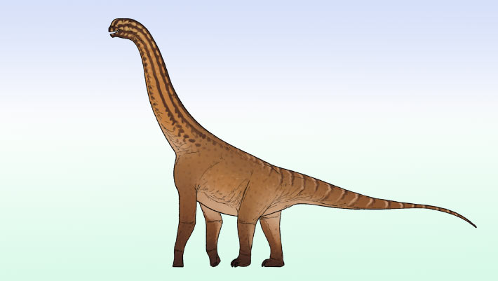
Small Sauropod Dinosaur from Jurassic Period Discovered in India
pelvis India Jurassic Middle Jurassic Dinosauria partial skeleton
Paleontologists in India have unearthed a partial skeleton of a diminutive, previously unknown neosauropod dinosaur from the Middle Jurassic rocks of Gujarat’s Kachchh Basin. The post Small Sauropod Dinosaur from Jurassic Period Discovered in India appeared first on Sci.News: Breaking Science News.
21/07/2026 sci-news
New Four-winged Dinosaur from China Described
feather limb description China Cretaceous Early Cretaceous fossil Dinosauria Microraptorinae new species partial
Palaeontologists have described a new species of microraptorine theropod from north-western China. Named Jian changmaensis, this small, feathered dinosaur probably glided on four wings. The fossil provides fresh insights into Early Cretaceous ecosystems and extends the known range of the Microraptorinae. The fossil material consists of an articulated partial left pectoral girdle and forelimb. It
08/06/2026 everythingdinosaur
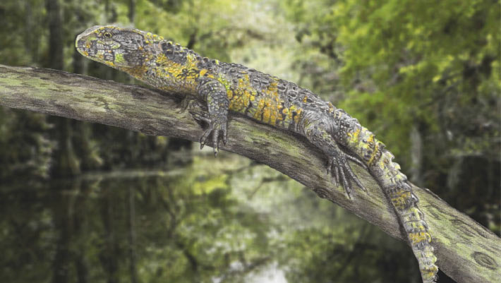
83-Million-Year-Old Crocodile Lizard Fossil Unearthed in France
jaw France fossil discovery new species partial
Paleontologists have identified a new genus and species of pan-shinisaur lizard from a partial upper jaw discovered in southern France, pushing the presence of its lineage in Europe back by at least 30 million years. The post 83-Million-Year-Old Crocodile Lizard Fossil Unearthed in France appeared first on Sci.News: Breaking Science News.
22/05/2026 sci-news
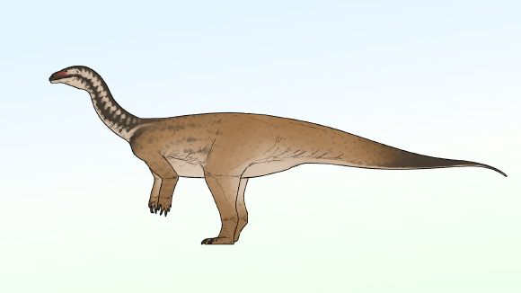
Early Sauropodomorph Dinosaur Unearthed in China
China Early Jurassic Jurassic Dinosauria Massopoda new species partial skeleton
A new genus and species of massopodan sauropodomorph that lived during the Early Jurassic epoch has been identified from a partial skeleton unearthed in southwestern China. The post Early Sauropodomorph Dinosaur Unearthed in China appeared first on Sci.News: Breaking Science News.
04/05/2026 sci-news
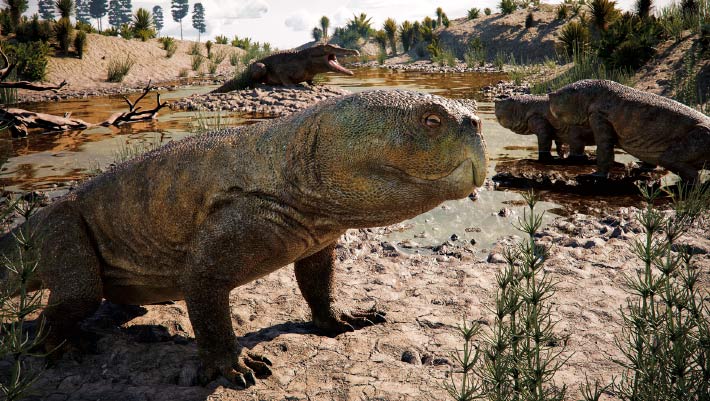
New Fossil from Brazil Reveals Unexpected Diversity among Pre-Dinosaur Herbivores
jaw Brazil Triassic fossil Dinosauria new species partial skull
Paleontologists in Brazil have identified a new genus and species of hyperodapedontine rhynchosaur -- an extinct lineage of beaked, herbivorous reptiles -- based on a partial skull and lower jaws recovered from Triassic rocks. The post New Fossil from Brazil Reveals Unexpected Diversity among Pre-Dinosaur Herbivores appeared first on Sci.News: Breaking Science News.
23/04/2026 sci-news