←

crest

Anatomical part

34 image(s) · 5 News

Image gallery

Rhomboid distal nasal process of the crest of Tsintaosaurus spinorhinus.

A. Partial distal nasal process of IVPP V725 in right lateral view. B. Lateral view of composite skull (reversed). C. partial premaxillonasal complex of IVPP V829 in right lateral view. D. Line drawing of (C) showing nasal-premaxilla articulation, and the displacement (arrow) experienced by the distal nasal process relative to its articular position. E. Mounted holotype skull (IVPP V725) of T. spinorhinus in left lateral view (reversed). F. Partial right distal nasal process (although catalogued as IVPP V725, this element corresponds to a different specimen than the type; reversed).
Taxa Tsintaosaurus

Rhomboid distal nasal process of the crest of Tsintaosaurus spinorhinus. A. Partial distal nasal process of IVPP V725 in right lateral view. B. Lateral view of composite skull (reversed). C. partial premaxillonasal complex of IVPP V829 in right lateral view. D. Line drawing of (C) showing nasal-premaxilla articulation, and the displacement (arrow) experienced by the distal nasal process relative to its articular position. E. Mounted holotype skull (IVPP V725) of T. spinorhinus in left lateral view (reversed). F. Partial right distal nasal process (although catalogued as IVPP V725, this element corresponds to a different specimen than the type; reversed).

crest drawing holotype specimen +3
Tachiraptor admirabilis gen et sp. nov. Holotype right tibia (IVIC-P-2867) in (a) lateral (proximal portion), (b) proximal, (c) lateral, (d) distal and (e) cranial (distal portion) views. Referred left ischium (IVIC-P-2868) in (f) lateral view. Abbreviations: ab, astragalar buttress; cc, cnemial crest; ai, cranial emargination; cn, caudal notch; fc, fibular condyle; fcr, fibular crest; ia, iliac articulation; in, intercondylar notch; it, incisura tibialis; keg, ‘knee extensor groove’; lg, longitudinal groove; lk, lateral kink; mc, medial condyle; om, outer malleolus; op, obturator plate; pp, pubic peduncle; pvr, ‘postero-ventral ridge’; rd, ridge; tn, tibial notch; vr, ventral ridge.
Taxa Tachiraptor

Tachiraptor admirabilis gen et sp. nov. Holotype right tibia (IVIC-P-2867) in (a) lateral (proximal portion), (b) proximal, (c) lateral, (d) distal and (e) cranial (distal portion) views. Referred left ischium (IVIC-P-2868) in (f) lateral view. Abbreviations: ab, astragalar buttress; cc, cnemial crest; ai, cranial emargination; cn, caudal notch; fc, fibular condyle; fcr, fibular crest; ia, iliac articulation; in, intercondylar notch; it, incisura tibialis; keg, ‘knee extensor groove’; lg, longitudinal groove; lk, lateral kink; mc, medial condyle; om, outer malleolus; op, obturator plate; pp, pubic peduncle; pvr, ‘postero-ventral ridge’; rd, ridge; tn, tibial notch; vr, ventral ridge.

crest holotype Averostra Tachiraptor
Photograph (A) and line drawings (B) of Gladocephaloideus jingangshanensis (JPM 2014–004). Abbreviations: ca, carpals; cr, coracoids; cv, cervical vertebrae; d, dentray; dg, deep groove along the mid-line of the mandibular symphysis; dv, dorsal vertebrae; dr, dorsal ribs; etp, extensor tendon process; f, frontal; fc, fifth carpal; fe, femur; fi, fibula; fin, fingers; h, humerus; il, ilium; m, maxilla; mmttsI-IV, metatrals I-IV; mttv, metatarsal V; nao, nasoantorbital opening; or, orbital; pcr, parietal crest; pm, premaxilla; pt, pteroid; ra, radius; rdl, radiale; sc, scapula; st, sternum; sl, sclerotic rings; t, teeth; tc, tooth sockets; ti, tibia; ul, ulna; wm, wing metacarpal; wph1-4, wing phalanges 1–4. Scale bar = 5 cm.
Taxa Gladocephaloideus

Photograph (A) and line drawings (B) of Gladocephaloideus jingangshanensis (JPM 2014–004). Abbreviations: ca, carpals; cr, coracoids; cv, cervical vertebrae; d, dentray; dg, deep groove along the mid-line of the mandibular symphysis; dv, dorsal vertebrae; dr, dorsal ribs; etp, extensor tendon process; f, frontal; fc, fifth carpal; fe, femur; fi, fibula; fin, fingers; h, humerus; il, ilium; m, maxilla; mmttsI-IV, metatrals I-IV; mttv, metatarsal V; nao, nasoantorbital opening; or, orbital; pcr, parietal crest; pm, premaxilla; pt, pteroid; ra, radius; rdl, radiale; sc, scapula; st, sternum; sl, sclerotic rings; t, teeth; tc, tooth sockets; ti, tibia; ul, ulna; wm, wing metacarpal; wph1-4, wing phalanges 1–4. Scale bar = 5 cm.

crest humerus scale tooth +2
Aralazhdarcho bostobensis, ZIN PH 57/43, a proximal fragment of a left humerus in proximal (a), ventral (b), anterior (c), dorsal (d), and posterior (e) views. This specimen is from the Shakh Shakh II locality in the northeasten Aral Sea region of Kazakhstan; Bostobe Formation, Upper Cretaceous (Santonian – lower Campanian). Abbreviations: h, humeral head; pf, pneumatic foramen; uc, ulnar crest. Scale bar is 10 mm.
Taxa Aralazhdarcho

Aralazhdarcho bostobensis, ZIN PH 57/43, a proximal fragment of a left humerus in proximal (a), ventral (b), anterior (c), dorsal (d), and posterior (e) views. This specimen is from the Shakh Shakh II locality in the northeasten Aral Sea region of Kazakhstan; Bostobe Formation, Upper Cretaceous (Santonian – lower Campanian). Abbreviations: h, humeral head; pf, pneumatic foramen; uc, ulnar crest. Scale bar is 10 mm.

crest humerus scale Kazakhstan +7
Skull of Caiuajara dobruskii gen. et sp. nov. (holotype, CP.V 1449) with the shape of an adult individual.

Scale bar equals 50: d, dentary; dcr, dentary crest; dep, depression; exp, ventral expansion of the premaxilla; f, frontal; fcr, frontal crest; fo, foraminae; m, maxilla; oc, occipital condyle; op, opisthotic; p, parietal; pm, premaxilla; pmcr, premaxillary crest; q, quadrate; soc, supraoccipital. The quadrate is inverted.
Taxa Caiuajara

Skull of Caiuajara dobruskii gen. et sp. nov. (holotype, CP.V 1449) with the shape of an adult individual. Scale bar equals 50: d, dentary; dcr, dentary crest; dep, depression; exp, ventral expansion of the premaxilla; f, frontal; fcr, frontal crest; fo, foraminae; m, maxilla; oc, occipital condyle; op, opisthotic; p, parietal; pm, premaxilla; pmcr, premaxillary crest; q, quadrate; soc, supraoccipital. The quadrate is inverted.

crest scale holotype Caiuajara +1
FSAC-OB 7, holotype right humerus, Simurghia robustus.
In (A), dorsal view, (B) ventral view, and (C) posterior view. Abbreviations: dpc, deltopectoral crest; msc, muscle scar; scpr, supracondylar process of the ectepicondyle; vp, ventral pillar; vt, ventral tubercle of the deltopectoral crest.
Taxa Simurghia

FSAC-OB 7, holotype right humerus, Simurghia robustus. In (A), dorsal view, (B) ventral view, and (C) posterior view. Abbreviations: dpc, deltopectoral crest; msc, muscle scar; scpr, supracondylar process of the ectepicondyle; vp, ventral pillar; vt, ventral tubercle of the deltopectoral crest.

crest humerus holotype Simurghia
FSAC-OB 232, holotype skeleton of Barbaridactylus grandis.
(A) Left humerus in anterior view, (B) right femur in anterior view, (C) right radius and ulna in posterior view, (D) cervical vertebra in ventral view, (E) left scapulocoracoid in medial view, and (F) posterior ramus of the right mandible in medial view.

Abbreviations: co, coracoid; cot, cotyle of mandible; cvr, cervical ribs; dm, dorsal margin of mandible; dpc, deltopectoral crest; gl, glenoid; fh, femoral head; gt, greater trochanter; hh, humeral head; hyp, hypapophysis; olp, olecranon process; pex, postexapophysis; pn, pneumatopore; rad, radius; sca, scapula; scp, supracondylar process of the ectepicondyle; uc, ulnar crest; uln, ulna; vm, ventral margin of mandible.
Taxa Barbaridactylus

FSAC-OB 232, holotype skeleton of Barbaridactylus grandis. (A) Left humerus in anterior view, (B) right femur in anterior view, (C) right radius and ulna in posterior view, (D) cervical vertebra in ventral view, (E) left scapulocoracoid in medial view, and (F) posterior ramus of the right mandible in medial view. Abbreviations: co, coracoid; cot, cotyle of mandible; cvr, cervical ribs; dm, dorsal margin of mandible; dpc, deltopectoral crest; gl, glenoid; fh, femoral head; gt, greater trochanter; hh, humeral head; hyp, hypapophysis; olp, olecranon process; pex, postexapophysis; pn, pneumatopore; rad, radius; sca, scapula; scp, supracondylar process of the ectepicondyle; uc, ulnar crest; uln, ulna; vm, ventral margin of mandible.

crest humerus vertebra holotype +2
Holotype of Jakapil kaniukura (MPCA-PV-630), postcranial bones. Speculative silhouette showing preserved elements (a); osteoderm distribution is speculative and partial to show non-osteodermal elements); dorsal vertebra elements in dorsal (b), right lateral (c) and anterior (d,e) views; sacral vertebra in left lateral view (f); mid-caudal vertebra in left lateral view (g); fragment of the mid-shaft of a dorsal rib in posterior view (the enlarged, broken posterior edge is highlighted (h); expanded distal ends of two dorsal ribs (i); left scapula in lateral view (j); right scapula in lateral view (k); right coracoid in lateral view (l); left and right humeri in anterior view (m); probable right ulna in lateral view (n); metacarpals, non-ungual and ungual phalanx in dorsal views (o); left femur elements in anterior view (p); proximal end of the right fibula in lateral view (q); distal end of the left tibia in anterior view (r); ischial elements in side view (s); cervical osteoderms in dorsal view (t), flat scutes in dorsal view (u), spine-like osteoderm in side view (v) and ossicle in dorsal view (w). ac acromial crest, aco asymmetrical cervical osteoderm, alp anterolateral process, ap acromial process, at anterior trochanter, bb basal bone, ebr expanded broken rib edge, di diapophysis, dpc deltopectoral crest, ft fourth trochanter, gl glenoid, mc metacarpals, nc neural canal, ncs neurocentral suture, ph non-ungual phalanx, pp pubic peduncle, poz postzygapophyses, rug marginal rugosities, sb scapular blade, sc scute, tp transverse process, uph ungual phalanx.
Taxa Jakapil

Holotype of Jakapil kaniukura (MPCA-PV-630), postcranial bones. Speculative silhouette showing preserved elements (a); osteoderm distribution is speculative and partial to show non-osteodermal elements); dorsal vertebra elements in dorsal (b), right lateral (c) and anterior (d,e) views; sacral vertebra in left lateral view (f); mid-caudal vertebra in left lateral view (g); fragment of the mid-shaft of a dorsal rib in posterior view (the enlarged, broken posterior edge is highlighted (h); expanded distal ends of two dorsal ribs (i); left scapula in lateral view (j); right scapula in lateral view (k); right coracoid in lateral view (l); left and right humeri in anterior view (m); probable right ulna in lateral view (n); metacarpals, non-ungual and ungual phalanx in dorsal views (o); left femur elements in anterior view (p); proximal end of the right fibula in lateral view (q); distal end of the left tibia in anterior view (r); ischial elements in side view (s); cervical osteoderms in dorsal view (t), flat scutes in dorsal view (u), spine-like osteoderm in side view (v) and ossicle in dorsal view (w). ac acromial crest, aco asymmetrical cervical osteoderm, alp anterolateral process, ap acromial process, at anterior trochanter, bb basal bone, ebr expanded broken rib edge, di diapophysis, dpc deltopectoral crest, ft fourth trochanter, gl glenoid, mc metacarpals, nc neural canal, ncs neurocentral suture, ph non-ungual phalanx, pp pubic peduncle, poz postzygapophyses, rug marginal rugosities, sb scapular blade, sc scute, tp transverse process, uph ungual phalanx.

bone crest vertebra holotype +2
Caihong juji holotype specimen (PMoL-B00175). Photographs of the slab (a) and counter slab (b) and line drawing (c) of the specimen based on both slabs. Photograph (d) and line drawing (e) of a composite of the rostrum of the skull and mandible exposed on the counter slab and the post-rostrum cranium exposed on the slab. Arrows indicate lacrimal crests. Question mark indicates uncertain identification. Scale bars: 10 cm a–c, 1 cm d and e. aof antorbital fenestra, cav caudal vertebra, cev cervical vertebra, dr dorsal rib, dv dorsal vertebra, ect ectopterygoid, emf external mandibular fenestra, en external naris, f feather, fu furcula, ga gastralia, hy hyoid, il ilium, is ischium, la left angular, lar left articular, lc left coracoid, lcr lacrimal crest, ld left dentary, lf left, frontal, lfe left femur, lh left humerus, lj left jugal, ll left lacrimal, lma left maxilla, lm left manus, ln left nasal, lp left pes, lpa left palatine, lpo left postorbital, lq left quadrate, lqj left quadratojugal, lr left radius, ls left scapula, lsp left splenial, lsa left surangular, lsq left squamosal, lt left tibiotarsus, lu left ulna, ma mandible, mf maxillary fenestra, o orbit, p parietal, pm premaxilla, pt pterygoid, pu pubis, rar right articular, rc right coracoid, rd right dentary, rfe right femur, rh right humerus, rm right manus, rp right pes, rpra right prearticular, rq right quadrate, rr right radius, rs right scapula, rt right tibiotarsus, ru right ulna, scl sclerotic bones, sk skull, sy synsacrum
Taxa Caihong

Caihong juji holotype specimen (PMoL-B00175). Photographs of the slab (a) and counter slab (b) and line drawing (c) of the specimen based on both slabs. Photograph (d) and line drawing (e) of a composite of the rostrum of the skull and mandible exposed on the counter slab and the post-rostrum cranium exposed on the slab. Arrows indicate lacrimal crests. Question mark indicates uncertain identification. Scale bars: 10 cm a–c, 1 cm d and e. aof antorbital fenestra, cav caudal vertebra, cev cervical vertebra, dr dorsal rib, dv dorsal vertebra, ect ectopterygoid, emf external mandibular fenestra, en external naris, f feather, fu furcula, ga gastralia, hy hyoid, il ilium, is ischium, la left angular, lar left articular, lc left coracoid, lcr lacrimal crest, ld left dentary, lf left, frontal, lfe left femur, lh left humerus, lj left jugal, ll left lacrimal, lma left maxilla, lm left manus, ln left nasal, lp left pes, lpa left palatine, lpo left postorbital, lq left quadrate, lqj left quadratojugal, lr left radius, ls left scapula, lsp left splenial, lsa left surangular, lsq left squamosal, lt left tibiotarsus, lu left ulna, ma mandible, mf maxillary fenestra, o orbit, p parietal, pm premaxilla, pt pterygoid, pu pubis, rar right articular, rc right coracoid, rd right dentary, rfe right femur, rh right humerus, rm right manus, rp right pes, rpra right prearticular, rq right quadrate, rr right radius, rs right scapula, rt right tibiotarsus, ru right ulna, scl sclerotic bones, sk skull, sy synsacrum

bone crest feather humerus +8
Left humerus of Haestasaurus becklesii (NHMUK R1870). A, anterior view; B, posterior view; Abbreviations: af, anconeal fossa; dp, deltopectoral crest; hh, humeral head; ltf, lateral triceps fossa; mtf, medial triceps fossa.

This fossil was found in strata of the Hastings Beds (late Berriasian—Valanginian in age) on the coast near Hastings, East Sussex, England.
Taxa Haestasaurus

Left humerus of Haestasaurus becklesii (NHMUK R1870). A, anterior view; B, posterior view; Abbreviations: af, anconeal fossa; dp, deltopectoral crest; hh, humeral head; ltf, lateral triceps fossa; mtf, medial triceps fossa. This fossil was found in strata of the Hastings Beds (late Berriasian—Valanginian in age) on the coast near Hastings, East Sussex, England.

crest humerus Berriasian Valanginian +2
Partial rostrum of Iberodactylus andreui gen. et sp. nov. (MPZ-2014/1). Photographic images (A–F)
and 3D renderings obtained from μCT data (G–L) in right lateral (A,G), left lateral (B,H), dorsal (C,I), palatal
(D,J), caudal (E,K), and cranial (F,L) views. Scale bar in cm. Abbreviations. pm: premaxilla; pmcr: premaxillary

crest; prid: palatal ridge; te: teeth; trb: trabeculae.
Taxa Iberodactylus

Partial rostrum of Iberodactylus andreui gen. et sp. nov. (MPZ-2014/1). Photographic images (A–F) and 3D renderings obtained from μCT data (G–L) in right lateral (A,G), left lateral (B,H), dorsal (C,I), palatal (D,J), caudal (E,K), and cranial (F,L) views. Scale bar in cm. Abbreviations. pm: premaxilla; pmcr: premaxillary crest; prid: palatal ridge; te: teeth; trb: trabeculae.

crest scale Iberodactylus partial
Holotype right pes of Vespersaurus paranaensis gen. et sp. nov. (MPCO.V 0065d1). (a) Distal end
of metatarsal II-IV in dorsal view. (b) Distal part of digit II in lateral view. (c) Distal part of digit IV in lateral
view. (d) Entire foot as preserved in lateral view. Anatomical abbreviations: ed, extensor depression; fd, flexor
depression; lcp, lateral collateral pit; mc, lateral crest; plg, proximolateral groove; vg, vertical groove; vlg, ventral

lateral groove; vm, ventral margin.
Taxa Vespersaurus

Holotype right pes of Vespersaurus paranaensis gen. et sp. nov. (MPCO.V 0065d1). (a) Distal end of metatarsal II-IV in dorsal view. (b) Distal part of digit II in lateral view. (c) Distal part of digit IV in lateral view. (d) Entire foot as preserved in lateral view. Anatomical abbreviations: ed, extensor depression; fd, flexor depression; lcp, lateral collateral pit; mc, lateral crest; plg, proximolateral groove; vg, vertical groove; vlg, ventral lateral groove; vm, ventral margin.

crest holotype Vespersaurus
Right femur of Meroktenos, MNHN.F.LES16c.

(A) Anterior, (B) medial, (C) posterior, (D) lateral, (E) proximal, and (F) distal views. ef, extensor fossa; fc, fibular condyle; fh, femoral head; lc, lateral condyle; lt, lesser trochanter; mc, medial condyle; pf, popliteal fossa; tfc, tibiofibular crest; 4t, fourth trochanter. (Photo credit: L Cazes.)
Taxa Meroktenos

Right femur of Meroktenos, MNHN.F.LES16c. (A) Anterior, (B) medial, (C) posterior, (D) lateral, (E) proximal, and (F) distal views. ef, extensor fossa; fc, fibular condyle; fh, femoral head; lc, lateral condyle; lt, lesser trochanter; mc, medial condyle; pf, popliteal fossa; tfc, tibiofibular crest; 4t, fourth trochanter. (Photo credit: L Cazes.)

crest Meroktenos
Skeletal reconstruction and postcranial anatomy of Asfaltovenator vialidadi, MPEF PV 3440. Centre: body outline with preserved elements indicated. (A) articulated cervical vertebrae three to five. (B) cervical vertebra 7. (C) articulated dorsal vertebrae four to seven (better preserved right side, reversed). (D) right humerus in anterior view. (E) right radius and ulna, medial view. (F) right manus, metacarpus in dorsal and digits in lateral view. (G) articulated proximal ends of right tibia and fibula in lateral and proximal views. Abbreviations: ag, anterior groove; cc, cnemial crest; cr, cervical rib; dc, distal carpal; di, diapophysis; dpc, deltpectoral crest; ec, ectepicondyle; ent, entepicondyle; epi, epipophysis; fi, fibula; fic, fibular condyle; hy, hyposphene; im, intermedium; it, internal tuberosity; lr, lateral ridge; mc, metacarpal; ns, neural spine; ol, olecranon; pa, parapophysis; pl, pleurocoel; poz, postzygapophysis; ppdl, paradiapophyseal lamina; prz, prezygapophysis; ra, radial. Scale bars are 100 cm (skeletal reconstruction), 5 cm (A–C) and 10 cm (D–G).
Taxa Asfaltovenator

Skeletal reconstruction and postcranial anatomy of Asfaltovenator vialidadi, MPEF PV 3440. Centre: body outline with preserved elements indicated. (A) articulated cervical vertebrae three to five. (B) cervical vertebra 7. (C) articulated dorsal vertebrae four to seven (better preserved right side, reversed). (D) right humerus in anterior view. (E) right radius and ulna, medial view. (F) right manus, metacarpus in dorsal and digits in lateral view. (G) articulated proximal ends of right tibia and fibula in lateral and proximal views. Abbreviations: ag, anterior groove; cc, cnemial crest; cr, cervical rib; dc, distal carpal; di, diapophysis; dpc, deltpectoral crest; ec, ectepicondyle; ent, entepicondyle; epi, epipophysis; fi, fibula; fic, fibular condyle; hy, hyposphene; im, intermedium; it, internal tuberosity; lr, lateral ridge; mc, metacarpal; ns, neural spine; ol, olecranon; pa, parapophysis; pl, pleurocoel; poz, postzygapophysis; ppdl, paradiapophyseal lamina; prz, prezygapophysis; ra, radial. Scale bars are 100 cm (skeletal reconstruction), 5 cm (A–C) and 10 cm (D–G).

crest humerus scale vertebra +3
Cuspicephalus scarfi MJMG K1918. A, Original skull on slab of mudstone, Lacking the mandible and dentition; B, outline diagram of preserved bone. Light grey is bone, dark grey is fibrous bone of sagittal crest, black is dental alveoli where unambiguous; C restoration of skull outline, with hypothetical lower jaw. Scale bar = 50 mm. Abbreviation: j, jugal; naof, nasoantorbital fenestra; m, maxilla; o, orbit; pm, premaxilla; q, quadrate; sc,

sagittal crest; sq, squamosal; tr np, trace of nasal process.
Taxa Cuspicephalus

Cuspicephalus scarfi MJMG K1918. A, Original skull on slab of mudstone, Lacking the mandible and dentition; B, outline diagram of preserved bone. Light grey is bone, dark grey is fibrous bone of sagittal crest, black is dental alveoli where unambiguous; C restoration of skull outline, with hypothetical lower jaw. Scale bar = 50 mm. Abbreviation: j, jugal; naof, nasoantorbital fenestra; m, maxilla; o, orbit; pm, premaxilla; q, quadrate; sc, sagittal crest; sq, squamosal; tr np, trace of nasal process.

bone crest scale Cuspicephalus +1
The holotype skeleton of Kamuysaurus japonicus gen. et sp. nov. (b) Reconstructed skeleton, showing recovered elements. Selected postcranial elements: cervical vertebrae (atlas, axis, and fourth and twelfth cervicals) in left lateral view (c), dorsal vertebrae (first, seventh, and sixteenth dorsals) in left lateral view (d), caudal vertebrae (anterior, middle, and posterior caudals) in left lateral view (e), left scapula (f) and coracoid (g) in lateral view, right sternum in ventral view (h), left humerus in anterior view (i), right ulna and radius in medial view (j), right manus in dorsal view (k), right pelvis in lateral view (l), right femur in anterior view (m), right tibia in anterior view (n), right fibula in lateral view (o), right astragalus and calcaneum, articulated positioned with tibia (p), and right pes in dorsal view (q). All scales are 10 cm except 1 m scale for (b). Abbreviations; ac, acromion process; ast, astragalus; cal, calcaneum; dpc, deltopectoral crest; fi, fibula; icg, intercondylar groove; il, ilium; is, ischium; ltr, lesser trochanter; mc2, metacarpal II; mc5, metacarpal V; mt2, metatarsal II; mt4, metatarsal IV; nc, neurocentrum; od, odontoid; olp, olecranon process; pc, pleurocentrum; pu, pubis; ra, radius; ti, tibia; ul, ulna; vp, ventral process.
Taxa Kamuysaurus

The holotype skeleton of Kamuysaurus japonicus gen. et sp. nov. (b) Reconstructed skeleton, showing recovered elements. Selected postcranial elements: cervical vertebrae (atlas, axis, and fourth and twelfth cervicals) in left lateral view (c), dorsal vertebrae (first, seventh, and sixteenth dorsals) in left lateral view (d), caudal vertebrae (anterior, middle, and posterior caudals) in left lateral view (e), left scapula (f) and coracoid (g) in lateral view, right sternum in ventral view (h), left humerus in anterior view (i), right ulna and radius in medial view (j), right manus in dorsal view (k), right pelvis in lateral view (l), right femur in anterior view (m), right tibia in anterior view (n), right fibula in lateral view (o), right astragalus and calcaneum, articulated positioned with tibia (p), and right pes in dorsal view (q). All scales are 10 cm except 1 m scale for (b). Abbreviations; ac, acromion process; ast, astragalus; cal, calcaneum; dpc, deltopectoral crest; fi, fibula; icg, intercondylar groove; il, ilium; is, ischium; ltr, lesser trochanter; mc2, metacarpal II; mc5, metacarpal V; mt2, metatarsal II; mt4, metatarsal IV; nc, neurocentrum; od, odontoid; olp, olecranon process; pc, pleurocentrum; pu, pubis; ra, radius; ti, tibia; ul, ulna; vp, ventral process.

crest humerus pelvis scale +3
1 2 3

News

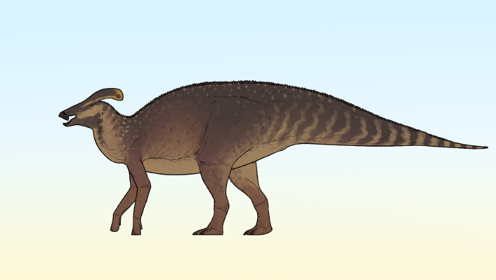
New Crested Dinosaur Species Identified in Canada
crest Canada Campanian Cretaceous Dinosauria Hadrosauridae dating new species
A new genus and species of lambeosaurine hadrosaurid dinosaur has been identified in southern Alberta, Canada, dating back 77 million years to the Campanian age of the Cretaceous period. The post New Crested Dinosaur Species Identified in Canada appeared first on Sci.News: Breaking Science News.
14/07/2026 sci-news
A giant blade-crested spinosaurus, the “hell heron,” discovered in the Sahara
crest predator Niger fossil Dinosauria Spinosauria
Deep in the heart of the Sahara, scientists have uncovered Spinosaurus mirabilis — a spectacular new predator crowned with a massive, scimitar-shaped crest that may once have blazed with color under the desert sun. Discovered in remote inland river deposits in Niger, the fossil rewrites what we thought we knew about spinosaur dinosaurs, suggesting they weren’t fully aquatic hunters but powerful waders stalking fish in forested waterways hundreds of miles from the sea.
23/02/2026 sciencedaily
Corythosaurus: Beast of the Week
crest Canada Canardia Corythosaurus Dinosauria Hadrosauria Hadrosauridae Hypacrosaurus Lambeosauria Parasaurolophus Velafrons
This week we will take a look at a well known duckbill dinosaur.  Enter Corythosaurus!  Corythosaurus was a plant eater that belonged to the lambiosaurine group within the family, Hadrosauridae. (duckbills)  Lambiosaurine hadrosaurs typically had hollow crests on their heads and narrower beaks compared to other kinds hadrosaurs.  It was closely related to other lambiosaurine hadrosaurs such as Parasaurolophus, Hypacrosaurus, Lambeosaurus, and Velafrons.  It lived in what is now Alberta, Canada,
15/02/2026 prehistoricbeastoftheweek
Parasaurolophus: Beast of the Week
crest Cretaceous Late Cretaceous specimen Canardia Dinosauria Parasaurolophus
This week we will be taking a look at a very popular duck-billed dinosaur.  Say hello to Parasaurolophus!  Parasaurolophus was a plant eater that lived in what is now North America during the Late Cretaceous, about 77 to 73 million years ago.  Parasaurolophus measured about 30 feet (9.1 meters) long from beak to tail, but certain incomplete specimens show evidence of having been a bit larger.  Parasaurolophus is most well-known for its long, curved crest that grew from the back of its head, givi
21/12/2025 prehistoricbeastoftheweek
Dinosaur mummy found with hooves and a hidden crest
crest scale movie Dinosauria Edmontosaurus
Scientists have reconstructed the most complete and lifelike profile of Edmontosaurus annectens thanks to an extraordinary preservation process called clay templating, in which a thin clay film captured the dinosaur’s skin, scales, spikes, and even hooves in three dimensions. By combining newly excavated “mummies,” advanced imaging, and artistic reconstruction, researchers revealed a tall crest, a single row of tail spikes, delicate pebble-like scales, and—most remarkably—the earliest known hoov
30/11/2025 sciencedaily