tissue

Anatomical part

7 image(s) · 3 News

Image gallery

Life restoration of the mosasaurine mosasaurid Eremiasaurus, with unknown portions and soft tissues based primarily on Prognathodon and supplemented with Mosasaurus where needed.
References
Leblanc, A.R.H.; Caldwell, M.W.; Bardet, N. (2012). "A new mosasaurine from the Maastrichtian (Upper Cretaceous) phosphates of Morocco and its implications for mosasaurine systematics". Journal of Vertebrate Paleontology 32 (1): 82–104.
Lindgren, J.; Kaddumi, H.; Polcyn, M. (2013). "Soft tissue preservation in a fossil marine lizard with a bilobed tail fin". Nature Communications 4: 2423. DOI:10.1038/ncomms3423.
Konishi, T.; Brinkman, D.; Massare, J.A.; Caldwell, M.W. (2011). "New exceptional specimens of Prognathodon overtoni (Squamata, Mosasauridae) from the upper Campanian of Alberta, Canada, and the systematics and ecology of the genus". Journal of Vertebrate Paleontology 31 (5): 1026–1046.
Russell, D.A. (1967). "Systematics and morphology of American mosasaurs". Bulletin of the Peabody Museum of Natural History 23: 1–241.

Life restoration of the mosasaurine mosasaurid Eremiasaurus, with unknown portions and soft tissues based primarily on Prognathodon and supplemented with Mosasaurus where needed. References Leblanc, A.R.H.; Caldwell, M.W.; Bardet, N. (2012). "A new mosasaurine from the Maastrichtian (Upper Cretaceous) phosphates of Morocco and its implications for mosasaurine systematics". Journal of Vertebrate Paleontology 32 (1): 82–104. Lindgren, J.; Kaddumi, H.; Polcyn, M. (2013). "Soft tissue preservation in a fossil marine lizard with a bilobed tail fin". Nature Communications 4: 2423. DOI:10.1038/ncomms3423. Konishi, T.; Brinkman, D.; Massare, J.A.; Caldwell, M.W. (2011). "New exceptional specimens of Prognathodon overtoni (Squamata, Mosasauridae) from the upper Campanian of Alberta, Canada, and the systematics and ecology of the genus". Journal of Vertebrate Paleontology 31 (5): 1026–1046. Russell, D.A. (1967). "Systematics and morphology of American mosasaurs". Bulletin of the Peabody Museum of Natural History 23: 1–241.

tissue ecology museum Canada +11
Skeletal restoration of Anchiornis huxleyi by Scott Hartman (2017), incorporating the soft tissue outlines revealed by laser fluorescence studies.
Taxa Anchiornithidae

Skeletal restoration of Anchiornis huxleyi by Scott Hartman (2017), incorporating the soft tissue outlines revealed by laser fluorescence studies.

tissue Anchiornis Anchiornithidae
Holotype of Eosipterus yangi GMV 2117, displaying soft tissue preservation around the hind limbs and/or ankles.
Taxa Eosipterus

Holotype of Eosipterus yangi GMV 2117, displaying soft tissue preservation around the hind limbs and/or ankles.

limb tissue holotype Eosipterus
The National history museum in Milan, Italy. Fossil of a juvenile individual of Scipionyx samniticus. The fossil (to date the first of this species ever) preserves in an exceptional way clear traces of soft tissues - a rather rare event. Picture by Giovanni Dall'Orto, April 22 2007.
Taxa Scipionyx

The National history museum in Milan, Italy. Fossil of a juvenile individual of Scipionyx samniticus. The fossil (to date the first of this species ever) preserves in an exceptional way clear traces of soft tissues - a rather rare event. Picture by Giovanni Dall'Orto, April 22 2007.

tissue museum Italy fossil +2
Main evolutionary steps proposed for the morphofunctional and postural changes of the sauropod pedes. (A) Sauropod body mass through time (in metric tons) based on the sauropod body mass estimations of (41) (NB: data lacking for the second half of the Upper Cretaceous so illustrated here faded, in continuity with the data recorded in the Cretaceous). Schematic outlines of selected large specimens illustrated in the curve, including (from left to right) P. engelhardti, Vulcanodon karibaensis, R. brownei, G. brancai, Cedarosaurus weiskopfae, and Notocolossus gonzalezparejasi. (B) Projected evolutionary changes occurring in the sauropod pes associated with trend in body mass, including 1, skeletal and functional digitigrade pedal posture among basal non-sauropod sauropodomorphs with an incipient soft tissue pad (ISP) (see figs. S34 and S35); 2 and 3, expansion of a well-developed soft tissue pad beneath the elevated pedal bones (SP), resulting in a functionally plantigrade pes + retention of skeletal posture within a range of digitigrady; 4, retention of a soft tissue pad and yet undetermined trend toward more elevated bones; 5, conservation of the neomorphic soft tissue pad within all lineages. Selected examples of well-preserved non-sauropod sauropodomorph and sauropod pedal tracks illustrated above the trends, including (from left to right) Evazoum siriguii; Pseudotetrasauropus bipedoida, Eosauropus isp., Lavinipes cheminii; Kalosauropus pollex, Liujianpus shunan, Polyonyx gomesi; Parabrontopodus mcintoshi; Brontopodus birdi; Titanopodus mendozensis; and unnamed Asian sauropod track. Source of adapted drawing and notes are listed in table S9 and data S2.
Taxa Evazoum

Main evolutionary steps proposed for the morphofunctional and postural changes of the sauropod pedes. (A) Sauropod body mass through time (in metric tons) based on the sauropod body mass estimations of (41) (NB: data lacking for the second half of the Upper Cretaceous so illustrated here faded, in continuity with the data recorded in the Cretaceous). Schematic outlines of selected large specimens illustrated in the curve, including (from left to right) P. engelhardti, Vulcanodon karibaensis, R. brownei, G. brancai, Cedarosaurus weiskopfae, and Notocolossus gonzalezparejasi. (B) Projected evolutionary changes occurring in the sauropod pes associated with trend in body mass, including 1, skeletal and functional digitigrade pedal posture among basal non-sauropod sauropodomorphs with an incipient soft tissue pad (ISP) (see figs. S34 and S35); 2 and 3, expansion of a well-developed soft tissue pad beneath the elevated pedal bones (SP), resulting in a functionally plantigrade pes + retention of skeletal posture within a range of digitigrady; 4, retention of a soft tissue pad and yet undetermined trend toward more elevated bones; 5, conservation of the neomorphic soft tissue pad within all lineages. Selected examples of well-preserved non-sauropod sauropodomorph and sauropod pedal tracks illustrated above the trends, including (from left to right) Evazoum siriguii; Pseudotetrasauropus bipedoida, Eosauropus isp., Lavinipes cheminii; Kalosauropus pollex, Liujianpus shunan, Polyonyx gomesi; Parabrontopodus mcintoshi; Brontopodus birdi; Titanopodus mendozensis; and unnamed Asian sauropod track. Source of adapted drawing and notes are listed in table S9 and data S2.

bone tissue Cretaceous specimen +6
Main evolutionary steps proposed for the morphofunctional and postural changes of the sauropod pedes. (A) Sauropod body mass through time (in metric tons) based on the sauropod body mass estimations of (41) (NB: data lacking for the second half of the Upper Cretaceous so illustrated here faded, in continuity with the data recorded in the Cretaceous). Schematic outlines of selected large specimens illustrated in the curve, including (from left to right) P. engelhardti, Vulcanodon karibaensis, R. brownei, G. brancai, Cedarosaurus weiskopfae, and Notocolossus gonzalezparejasi. (B) Projected evolutionary changes occurring in the sauropod pes associated with trend in body mass, including 1, skeletal and functional digitigrade pedal posture among basal non-sauropod sauropodomorphs with an incipient soft tissue pad (ISP) (see figs. S34 and S35); 2 and 3, expansion of a well-developed soft tissue pad beneath the elevated pedal bones (SP), resulting in a functionally plantigrade pes + retention of skeletal posture within a range of digitigrady; 4, retention of a soft tissue pad and yet undetermined trend toward more elevated bones; 5, conservation of the neomorphic soft tissue pad within all lineages. Selected examples of well-preserved non-sauropod sauropodomorph and sauropod pedal tracks illustrated above the trends, including (from left to right) Evazoum siriguii; Pseudotetrasauropus bipedoida, Eosauropus isp., Lavinipes cheminii; Kalosauropus pollex, Liujianpus shunan, Polyonyx gomesi; Parabrontopodus mcintoshi; Brontopodus birdi; Titanopodus mendozensis; and unnamed Asian sauropod track. Source of adapted drawing and notes are listed in table S9 and data S2.
Taxa Kalosauropus

Main evolutionary steps proposed for the morphofunctional and postural changes of the sauropod pedes. (A) Sauropod body mass through time (in metric tons) based on the sauropod body mass estimations of (41) (NB: data lacking for the second half of the Upper Cretaceous so illustrated here faded, in continuity with the data recorded in the Cretaceous). Schematic outlines of selected large specimens illustrated in the curve, including (from left to right) P. engelhardti, Vulcanodon karibaensis, R. brownei, G. brancai, Cedarosaurus weiskopfae, and Notocolossus gonzalezparejasi. (B) Projected evolutionary changes occurring in the sauropod pes associated with trend in body mass, including 1, skeletal and functional digitigrade pedal posture among basal non-sauropod sauropodomorphs with an incipient soft tissue pad (ISP) (see figs. S34 and S35); 2 and 3, expansion of a well-developed soft tissue pad beneath the elevated pedal bones (SP), resulting in a functionally plantigrade pes + retention of skeletal posture within a range of digitigrady; 4, retention of a soft tissue pad and yet undetermined trend toward more elevated bones; 5, conservation of the neomorphic soft tissue pad within all lineages. Selected examples of well-preserved non-sauropod sauropodomorph and sauropod pedal tracks illustrated above the trends, including (from left to right) Evazoum siriguii; Pseudotetrasauropus bipedoida, Eosauropus isp., Lavinipes cheminii; Kalosauropus pollex, Liujianpus shunan, Polyonyx gomesi; Parabrontopodus mcintoshi; Brontopodus birdi; Titanopodus mendozensis; and unnamed Asian sauropod track. Source of adapted drawing and notes are listed in table S9 and data S2.

bone tissue Cretaceous specimen +6
New reconstruction of Tylosaurus proriger, based on recent data about mosasaur's soft tissue

New reconstruction of Tylosaurus proriger, based on recent data about mosasaur's soft tissue

tissue Tylosaurus
Life restoration of the mosasaurine mosasaurid Eremiasaurus, with unknown portions and soft tissues based primarily on Prognathodon and supplemented with Mosasaurus where needed.
References
Leblanc, A.R.H.; Caldwell, M.W.; Bardet, N. (2012). "A new mosasaurine from the Maastrichtian (Upper Cretaceous) phosphates of Morocco and its implications for mosasaurine systematics". Journal of Vertebrate Paleontology 32 (1): 82–104.
Lindgren, J.; Kaddumi, H.; Polcyn, M. (2013). "Soft tissue preservation in a fossil marine lizard with a bilobed tail fin". Nature Communications 4: 2423. DOI:10.1038/ncomms3423.
Konishi, T.; Brinkman, D.; Massare, J.A.; Caldwell, M.W. (2011). "New exceptional specimens of Prognathodon overtoni (Squamata, Mosasauridae) from the upper Campanian of Alberta, Canada, and the systematics and ecology of the genus". Journal of Vertebrate Paleontology 31 (5): 1026–1046.
Russell, D.A. (1967). "Systematics and morphology of American mosasaurs". Bulletin of the Peabody Museum of Natural History 23: 1–241.
Taxa Eremiasaurus

Life restoration of the mosasaurine mosasaurid Eremiasaurus, with unknown portions and soft tissues based primarily on Prognathodon and supplemented with Mosasaurus where needed. References Leblanc, A.R.H.; Caldwell, M.W.; Bardet, N. (2012). "A new mosasaurine from the Maastrichtian (Upper Cretaceous) phosphates of Morocco and its implications for mosasaurine systematics". Journal of Vertebrate Paleontology 32 (1): 82–104. Lindgren, J.; Kaddumi, H.; Polcyn, M. (2013). "Soft tissue preservation in a fossil marine lizard with a bilobed tail fin". Nature Communications 4: 2423. DOI:10.1038/ncomms3423. Konishi, T.; Brinkman, D.; Massare, J.A.; Caldwell, M.W. (2011). "New exceptional specimens of Prognathodon overtoni (Squamata, Mosasauridae) from the upper Campanian of Alberta, Canada, and the systematics and ecology of the genus". Journal of Vertebrate Paleontology 31 (5): 1026–1046. Russell, D.A. (1967). "Systematics and morphology of American mosasaurs". Bulletin of the Peabody Museum of Natural History 23: 1–241.

tissue ecology museum Canada +11

News

518-million-year-old creature reveals the origins of spider fangs
limb tissue fossil
A tiny 518-million-year-old sea creature has revealed the earliest known evidence of the structures that eventually became spiders’ fangs. Hidden inside the fossil were pincerlike limbs and preserved soft tissues that help explain how the ancestors of spiders and scorpions evolved into formidable hunters.
04/08/2026 sciencedaily-paleo
113-Million-Year-Old Pterosaur Fossil Reveals What Flying Reptiles Ate
tissue feeding Brazil fossil Anhangueridae Pterosauria fish
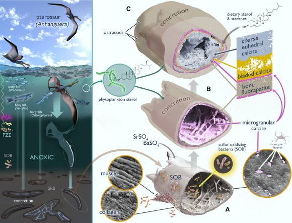
A 113-million-year-old pterosaur fossil from northeastern Brazil has yielded rare evidence of soft tissues, organic molecules and chemical traces of a diet heavy in fish and cephalopods such as squid or nautilus relatives. The post 113-Million-Year-Old Pterosaur Fossil Reveals What Flying Reptiles Ate appeared first on Sci.News: Breaking Science News.
19/06/2026 sci-news
Ground-breaking Study Strengthens the Case for Lips in Dinosaurs
skin tissue tooth Dinosauria biomechanics study
The debate about lips in dinosaurs continues.  Whether dinosaurs possessed extraoral tissue remains controversial. However, a remarkable new study has added substantial support to the idea that most dinosaurs possessed extraoral tissues that covered and protected their teeth. A new study, published in the journal "Palaeontology" provides compelling evidence that lips were the ancestral condition
15/06/2026 everythingdinosaur