←

vertèbre

Partie anatomique

70 image(s) · 12 Actualités

Galerie d'images

Preserved elements of Tataouinea hannibalis (ONM DT 1–48).
A, quarry map showing the orientation of collected elements. B, field photograph of the elements collected at the end of 2011, and C, of elements collected in 2013. Ca, caudal vertebra 1–17; S, sacral vertebra 1–5; r il, right ilium; l il, left ilium.
Taxons Tataouinea

Preserved elements of Tataouinea hannibalis (ONM DT 1–48). A, quarry map showing the orientation of collected elements. B, field photograph of the elements collected at the end of 2011, and C, of elements collected in 2013. Ca, caudal vertebra 1–17; S, sacral vertebra 1–5; r il, right ilium; l il, left ilium.

vertèbre Tataouinea
Holotype dorsal vertebra of Nopcsasopondylus.
Taxons Nopcsaspondylus

Holotype dorsal vertebra of Nopcsasopondylus.

vertèbre holotype Nopcsaspondylus
PLATE X X X I ~ r.
The vertebral column of Anoplosaurus c~rto~ot~s.
Fig. l. Left lateral aspect of third to seventh cervical eentrums, showing how
the bodies of the vertebrae increase in depth from before backward,
and indicating at the upper anterior corner of each an attachment
for a cervical rib.
1 a. Visceral aspect of third (?) cervical vertebra, showing flattened and
rounded under surface, and expansion towards the anterior end.
Fig. 2. Left lateral aspect of fourteenth to eighteenth eentrums of the series,
showing the somewhat compressed appearance of upper parts of the
bodies of the last five dorsal vertebra.
2 a. Visceral aspect of the last dorsal vertebra, 1~o. 18.
2 b. Posterior (?) articular end of the same centrum, No. 18.
3. The sacrum, showing the neural or superior surface, and giving evi-
dence of the great expansion of the spinal cord in the region of the
second and third sacral vertebra. The outlets for the escape of the
sacral nerves at the sides of the centrum are best seen in the first
three vertebrae.
4. Dorsal surface of vertebral end of a sacral rib, showing the massive end
for attachment to the sides of the centrums of the second and third
vertebrm. Its worn upper surface gave attachment to the neural
arch.
5. Left lateral aspect of a sequence of six early caudal vertebrm numbered
25 to 30, showing decreasing depth of the centrum posteriorly, and
diminishing size in the surface for the attachment of the sacral rib
which remains in union with the 30th vertebra. The chevron facet, e,
also diminishes in size.
5 a. Posterior articular surface and facet for the chevron bone of the

vertebra ~No. 26.
Taxons Anoplosaurus

PLATE X X X I ~ r. The vertebral column of Anoplosaurus c~rto~ot~s. Fig. l. Left lateral aspect of third to seventh cervical eentrums, showing how the bodies of the vertebrae increase in depth from before backward, and indicating at the upper anterior corner of each an attachment for a cervical rib. 1 a. Visceral aspect of third (?) cervical vertebra, showing flattened and rounded under surface, and expansion towards the anterior end. Fig. 2. Left lateral aspect of fourteenth to eighteenth eentrums of the series, showing the somewhat compressed appearance of upper parts of the bodies of the last five dorsal vertebra. 2 a. Visceral aspect of the last dorsal vertebra, 1~o. 18. 2 b. Posterior (?) articular end of the same centrum, No. 18. 3. The sacrum, showing the neural or superior surface, and giving evi- dence of the great expansion of the spinal cord in the region of the second and third sacral vertebra. The outlets for the escape of the sacral nerves at the sides of the centrum are best seen in the first three vertebrae. 4. Dorsal surface of vertebral end of a sacral rib, showing the massive end for attachment to the sides of the centrums of the second and third vertebrm. Its worn upper surface gave attachment to the neural arch. 5. Left lateral aspect of a sequence of six early caudal vertebrm numbered 25 to 30, showing decreasing depth of the centrum posteriorly, and diminishing size in the surface for the attachment of the sacral rib which remains in union with the 30th vertebra. The chevron facet, e, also diminishes in size. 5 a. Posterior articular surface and facet for the chevron bone of the vertebra ~No. 26.

os vertèbre Anoplosaurus
Vertebra of the rebbachisaurid sauropod Histriasaurus from Croatia
Taxons Histriasaurus

Vertebra of the rebbachisaurid sauropod Histriasaurus from Croatia

vertèbre Croatie Histriasaurus Rebbachisauridae
A photograph of the skeleton of Jianchangosaurus yixianensis gen. et sp. nov. (41HIII-0308A) (A) and line drawing (B).

Abbreviations: at, atlas; ax, axis; cav, caudal vertebra; cv, cervical vertebra; co, coracoid; dc, distal carpal; dv, dorsal vertebra; l., left; f, femur; fi, fibula; fur, furcula; hu, humerus; il, ilium; is, ischium; pu, pubis; ra, radius; sc, scapula; ti, tibia; ul, ulna; mc, metacarpal; mt, metatarsal; mxf, maxillary fenestra; pat, proatlas; r., right; sr, sacral rib; sv, sacral vertebra; ung, ungual. All elements of the skeleton are preserved except the distal half of the caudal vertebrae. Dashed lines of metatarsals indicate areas that have been reconstructed. The middle portion of the neck, from the fourth to ninth cervical vertebrae, and the pedal phalanges have been repositioned. The rest of elements of this specimen are in the original position.
Taxons Jianchangosaurus

A photograph of the skeleton of Jianchangosaurus yixianensis gen. et sp. nov. (41HIII-0308A) (A) and line drawing (B). Abbreviations: at, atlas; ax, axis; cav, caudal vertebra; cv, cervical vertebra; co, coracoid; dc, distal carpal; dv, dorsal vertebra; l., left; f, femur; fi, fibula; fur, furcula; hu, humerus; il, ilium; is, ischium; pu, pubis; ra, radius; sc, scapula; ti, tibia; ul, ulna; mc, metacarpal; mt, metatarsal; mxf, maxillary fenestra; pat, proatlas; r., right; sr, sacral rib; sv, sacral vertebra; ung, ungual. All elements of the skeleton are preserved except the distal half of the caudal vertebrae. Dashed lines of metatarsals indicate areas that have been reconstructed. The middle portion of the neck, from the fourth to ninth cervical vertebrae, and the pedal phalanges have been repositioned. The rest of elements of this specimen are in the original position.

humérus vertèbre dessin spécimen +2
Skeletal reconstruction of Maraapunisaurus fragillimus, with restored dorsal vertebra in posterior view. Scaled from Lavocatisaurus, Agustinia, Comahuesaurus, Amazonsaurus, & other basal rebbachisaurids, with neck extended allometrically following Stevens & Parrish (2006). Human silhouette is myself.
Taxons Comahuesaurus

Skeletal reconstruction of Maraapunisaurus fragillimus, with restored dorsal vertebra in posterior view. Scaled from Lavocatisaurus, Agustinia, Comahuesaurus, Amazonsaurus, & other basal rebbachisaurids, with neck extended allometrically following Stevens & Parrish (2006). Human silhouette is myself.

vertèbre Agustinia Amazonsaurus Comahuesaurus +3
FSAC-OB 232, holotype skeleton of Barbaridactylus grandis.
(A) Left humerus in anterior view, (B) right femur in anterior view, (C) right radius and ulna in posterior view, (D) cervical vertebra in ventral view, (E) left scapulocoracoid in medial view, and (F) posterior ramus of the right mandible in medial view.

Abbreviations: co, coracoid; cot, cotyle of mandible; cvr, cervical ribs; dm, dorsal margin of mandible; dpc, deltopectoral crest; gl, glenoid; fh, femoral head; gt, greater trochanter; hh, humeral head; hyp, hypapophysis; olp, olecranon process; pex, postexapophysis; pn, pneumatopore; rad, radius; sca, scapula; scp, supracondylar process of the ectepicondyle; uc, ulnar crest; uln, ulna; vm, ventral margin of mandible.
Taxons Barbaridactylus

FSAC-OB 232, holotype skeleton of Barbaridactylus grandis. (A) Left humerus in anterior view, (B) right femur in anterior view, (C) right radius and ulna in posterior view, (D) cervical vertebra in ventral view, (E) left scapulocoracoid in medial view, and (F) posterior ramus of the right mandible in medial view. Abbreviations: co, coracoid; cot, cotyle of mandible; cvr, cervical ribs; dm, dorsal margin of mandible; dpc, deltopectoral crest; gl, glenoid; fh, femoral head; gt, greater trochanter; hh, humeral head; hyp, hypapophysis; olp, olecranon process; pex, postexapophysis; pn, pneumatopore; rad, radius; sca, scapula; scp, supracondylar process of the ectepicondyle; uc, ulnar crest; uln, ulna; vm, ventral margin of mandible.

crête humérus vertèbre holotype +2
Nemicolopterus crypticus holotype (IVPP V-14377).

(A) Skeleton overview, and (B) schematic drawing. (C) Skull (right lateral view), and (D) schematic drawing. Abbreviations: cdv, caudal vertebrae; co, coracoid; cv, cervical vertebra; d1–d4, digits 1–4; f, frontal; fe, femur; h, humerus; hy, hyoid; il, illium; is, ischium; j, jugal; l, left; la, lacrimal; mand, mandible; mc, metacarpal; mt, metatarsal; naof, nasoantorbital fenestra; or, orbit; pa, parietal; pm, premaxilla; ph, phalanx; ti, tibia; r, right; sca, scapula. Scale bars: A–B, 100 mm; C–D, 5 mm.
Taxons Nemicolopterus

Nemicolopterus crypticus holotype (IVPP V-14377). (A) Skeleton overview, and (B) schematic drawing. (C) Skull (right lateral view), and (D) schematic drawing. Abbreviations: cdv, caudal vertebrae; co, coracoid; cv, cervical vertebra; d1–d4, digits 1–4; f, frontal; fe, femur; h, humerus; hy, hyoid; il, illium; is, ischium; j, jugal; l, left; la, lacrimal; mand, mandible; mc, metacarpal; mt, metatarsal; naof, nasoantorbital fenestra; or, orbit; pa, parietal; pm, premaxilla; ph, phalanx; ti, tibia; r, right; sca, scapula. Scale bars: A–B, 100 mm; C–D, 5 mm.

humérus écaille vertèbre dessin +4
Holotype of Jakapil kaniukura (MPCA-PV-630), postcranial bones. Speculative silhouette showing preserved elements (a); osteoderm distribution is speculative and partial to show non-osteodermal elements); dorsal vertebra elements in dorsal (b), right lateral (c) and anterior (d,e) views; sacral vertebra in left lateral view (f); mid-caudal vertebra in left lateral view (g); fragment of the mid-shaft of a dorsal rib in posterior view (the enlarged, broken posterior edge is highlighted (h); expanded distal ends of two dorsal ribs (i); left scapula in lateral view (j); right scapula in lateral view (k); right coracoid in lateral view (l); left and right humeri in anterior view (m); probable right ulna in lateral view (n); metacarpals, non-ungual and ungual phalanx in dorsal views (o); left femur elements in anterior view (p); proximal end of the right fibula in lateral view (q); distal end of the left tibia in anterior view (r); ischial elements in side view (s); cervical osteoderms in dorsal view (t), flat scutes in dorsal view (u), spine-like osteoderm in side view (v) and ossicle in dorsal view (w). ac acromial crest, aco asymmetrical cervical osteoderm, alp anterolateral process, ap acromial process, at anterior trochanter, bb basal bone, ebr expanded broken rib edge, di diapophysis, dpc deltopectoral crest, ft fourth trochanter, gl glenoid, mc metacarpals, nc neural canal, ncs neurocentral suture, ph non-ungual phalanx, pp pubic peduncle, poz postzygapophyses, rug marginal rugosities, sb scapular blade, sc scute, tp transverse process, uph ungual phalanx.
Taxons Jakapil

Holotype of Jakapil kaniukura (MPCA-PV-630), postcranial bones. Speculative silhouette showing preserved elements (a); osteoderm distribution is speculative and partial to show non-osteodermal elements); dorsal vertebra elements in dorsal (b), right lateral (c) and anterior (d,e) views; sacral vertebra in left lateral view (f); mid-caudal vertebra in left lateral view (g); fragment of the mid-shaft of a dorsal rib in posterior view (the enlarged, broken posterior edge is highlighted (h); expanded distal ends of two dorsal ribs (i); left scapula in lateral view (j); right scapula in lateral view (k); right coracoid in lateral view (l); left and right humeri in anterior view (m); probable right ulna in lateral view (n); metacarpals, non-ungual and ungual phalanx in dorsal views (o); left femur elements in anterior view (p); proximal end of the right fibula in lateral view (q); distal end of the left tibia in anterior view (r); ischial elements in side view (s); cervical osteoderms in dorsal view (t), flat scutes in dorsal view (u), spine-like osteoderm in side view (v) and ossicle in dorsal view (w). ac acromial crest, aco asymmetrical cervical osteoderm, alp anterolateral process, ap acromial process, at anterior trochanter, bb basal bone, ebr expanded broken rib edge, di diapophysis, dpc deltopectoral crest, ft fourth trochanter, gl glenoid, mc metacarpals, nc neural canal, ncs neurocentral suture, ph non-ungual phalanx, pp pubic peduncle, poz postzygapophyses, rug marginal rugosities, sb scapular blade, sc scute, tp transverse process, uph ungual phalanx.

os crête vertèbre holotype +2
Caihong juji holotype specimen (PMoL-B00175). Photographs of the slab (a) and counter slab (b) and line drawing (c) of the specimen based on both slabs. Photograph (d) and line drawing (e) of a composite of the rostrum of the skull and mandible exposed on the counter slab and the post-rostrum cranium exposed on the slab. Arrows indicate lacrimal crests. Question mark indicates uncertain identification. Scale bars: 10 cm a–c, 1 cm d and e. aof antorbital fenestra, cav caudal vertebra, cev cervical vertebra, dr dorsal rib, dv dorsal vertebra, ect ectopterygoid, emf external mandibular fenestra, en external naris, f feather, fu furcula, ga gastralia, hy hyoid, il ilium, is ischium, la left angular, lar left articular, lc left coracoid, lcr lacrimal crest, ld left dentary, lf left, frontal, lfe left femur, lh left humerus, lj left jugal, ll left lacrimal, lma left maxilla, lm left manus, ln left nasal, lp left pes, lpa left palatine, lpo left postorbital, lq left quadrate, lqj left quadratojugal, lr left radius, ls left scapula, lsp left splenial, lsa left surangular, lsq left squamosal, lt left tibiotarsus, lu left ulna, ma mandible, mf maxillary fenestra, o orbit, p parietal, pm premaxilla, pt pterygoid, pu pubis, rar right articular, rc right coracoid, rd right dentary, rfe right femur, rh right humerus, rm right manus, rp right pes, rpra right prearticular, rq right quadrate, rr right radius, rs right scapula, rt right tibiotarsus, ru right ulna, scl sclerotic bones, sk skull, sy synsacrum
Taxons Caihong

Caihong juji holotype specimen (PMoL-B00175). Photographs of the slab (a) and counter slab (b) and line drawing (c) of the specimen based on both slabs. Photograph (d) and line drawing (e) of a composite of the rostrum of the skull and mandible exposed on the counter slab and the post-rostrum cranium exposed on the slab. Arrows indicate lacrimal crests. Question mark indicates uncertain identification. Scale bars: 10 cm a–c, 1 cm d and e. aof antorbital fenestra, cav caudal vertebra, cev cervical vertebra, dr dorsal rib, dv dorsal vertebra, ect ectopterygoid, emf external mandibular fenestra, en external naris, f feather, fu furcula, ga gastralia, hy hyoid, il ilium, is ischium, la left angular, lar left articular, lc left coracoid, lcr lacrimal crest, ld left dentary, lf left, frontal, lfe left femur, lh left humerus, lj left jugal, ll left lacrimal, lma left maxilla, lm left manus, ln left nasal, lp left pes, lpa left palatine, lpo left postorbital, lq left quadrate, lqj left quadratojugal, lr left radius, ls left scapula, lsp left splenial, lsa left surangular, lsq left squamosal, lt left tibiotarsus, lu left ulna, ma mandible, mf maxillary fenestra, o orbit, p parietal, pm premaxilla, pt pterygoid, pu pubis, rar right articular, rc right coracoid, rd right dentary, rfe right femur, rh right humerus, rm right manus, rp right pes, rpra right prearticular, rq right quadrate, rr right radius, rs right scapula, rt right tibiotarsus, ru right ulna, scl sclerotic bones, sk skull, sy synsacrum

os crête plume humérus +8
Istiorachis macarthurae holotype (MIWG 6643). The anterior-most seven caudal vertebrae in left lateral view with neural spines reconstructed. Abbreviations: Cd, caudal vertebra; SC, sacrocaudal. Scale bar represents 50 mm.
Taxons Istiorachis

Istiorachis macarthurae holotype (MIWG 6643). The anterior-most seven caudal vertebrae in left lateral view with neural spines reconstructed. Abbreviations: Cd, caudal vertebra; SC, sacrocaudal. Scale bar represents 50 mm.

écaille vertèbre holotype Istiorachis
Identifier: annalsofmedicalh01pack (find matches)
Title: Annals of medical history
Year: 1917 (1910s)
Authors:  Packard, Francis R. (Francis Randolph), 1870-1950
Subjects:  Medicine
Publisher:  New York P.B. Hoeber
Contributing Library:  Gerstein - University of Toronto
Digitizing Sponsor:  University of Toronto

View Book Page: Book Viewer
About This Book: Catalog Entry
View All Images: All Images From Book
Click here to view book online to see this illustration in context in a browseable online version of this book.

Text Appearing Before Image:
ail strongly Hexed, and the toescontracted and appressed. The whole atti-tude strongly suggests a spastic distress,possibly brought on by some form ofpoisoning of the central nervous system,from infection or the deglutition of somepoisonous substance. 14. Osteomalacia is evidently the causeof the hypertrophy of the bones of Lim-nocyron potens, an early carnivore from theWashakie Eocene of Wyoming, nearly3,000,000 years old. MATERIALS AND METHODS The material described in the presentpaper has been loaned the writer lor de-scription by the Field Museum of Chii Osborn: Hull. Amcr. \lus. Natl. Hist., 1917,vol. 35. !>• 733. P>- 28. Studies in Paleopathology 393 by the American Museum of Natural His-tory of New York City, by Walker Museumof the University of Chicago, and by the Uni-versity of Kansas Natural History Museum. made by the well-known petrographic meth-ods so common in all geological labora-tories. The diagnoses, where they are at-tempted, arc made from comparisons of the
Text Appearing After Image:
Fig. 20. The skeleton of Strutbiomimus altus, a small dinosaur from the BeliyRiver series (Cretaceous), Red Deer River, Alberta, Canada, now regarded as ofapproximately the same age as the Judith River series. The unique feature ofthe skull is the total absence of teeth, with a size of skull one-third larger thanthe ostrich and a length of body of about fifteen feet. The position of the skeletonis decidedly that of the opisthotonos which may be regarded as an indication ofdisease. (After Osborn). A beautiful specimen of an osteoma, theonly one known so far, on the vertebra of aKansas Cretaceous mosasaur, was given thewriter by Dr. J. M. Armstrong of St. Paul.The writer expresses his obligations to thegentlemen connected with the above-men-tioned institutions and to Dr. Armstrong.The methods used are a combination ofprocedures in the various lines involved.Microscopic sections, which can be madethin enough for immersion lens study, are material with similar lesions in recent hu-man mat

Note About Images

Please note that these images are extracted from scanned page images that may have been digitally enhanced for readability - coloration and appearance of these illustrations may not perfectly resemble the original work.
Taxons Hexing

Identifier: annalsofmedicalh01pack (find matches) Title: Annals of medical history Year: 1917 (1910s) Authors: Packard, Francis R. (Francis Randolph), 1870-1950 Subjects: Medicine Publisher: New York P.B. Hoeber Contributing Library: Gerstein - University of Toronto Digitizing Sponsor: University of Toronto View Book Page: Book Viewer About This Book: Catalog Entry View All Images: All Images From Book Click here to view book online to see this illustration in context in a browseable online version of this book. Text Appearing Before Image: ail strongly Hexed, and the toescontracted and appressed. The whole atti-tude strongly suggests a spastic distress,possibly brought on by some form ofpoisoning of the central nervous system,from infection or the deglutition of somepoisonous substance. 14. Osteomalacia is evidently the causeof the hypertrophy of the bones of Lim-nocyron potens, an early carnivore from theWashakie Eocene of Wyoming, nearly3,000,000 years old. MATERIALS AND METHODS The material described in the presentpaper has been loaned the writer lor de-scription by the Field Museum of Chii Osborn: Hull. Amcr. \lus. Natl. Hist., 1917,vol. 35. !>• 733. P>- 28. Studies in Paleopathology 393 by the American Museum of Natural His-tory of New York City, by Walker Museumof the University of Chicago, and by the Uni-versity of Kansas Natural History Museum. made by the well-known petrographic meth-ods so common in all geological labora-tories. The diagnoses, where they are at-tempted, arc made from comparisons of the Text Appearing After Image: Fig. 20. The skeleton of Strutbiomimus altus, a small dinosaur from the BeliyRiver series (Cretaceous), Red Deer River, Alberta, Canada, now regarded as ofapproximately the same age as the Judith River series. The unique feature ofthe skull is the total absence of teeth, with a size of skull one-third larger thanthe ostrich and a length of body of about fifteen feet. The position of the skeletonis decidedly that of the opisthotonos which may be regarded as an indication ofdisease. (After Osborn). A beautiful specimen of an osteoma, theonly one known so far, on the vertebra of aKansas Cretaceous mosasaur, was given thewriter by Dr. J. M. Armstrong of St. Paul.The writer expresses his obligations to thegentlemen connected with the above-men-tioned institutions and to Dr. Armstrong.The methods used are a combination ofprocedures in the various lines involved.Microscopic sections, which can be madethin enough for immersion lens study, are material with similar lesions in recent hu-man mat Note About Images Please note that these images are extracted from scanned page images that may have been digitally enhanced for readability - coloration and appearance of these illustrations may not perfectly resemble the original work.

os vertèbre livre musée +11
Caudal vertebra of Astrophocaudia SMU 61732 photo from 2014-04-09 PM
Taxons Astrophocaudia

Caudal vertebra of Astrophocaudia SMU 61732 photo from 2014-04-09 PM

vertèbre Astrophocaudia
Alvarezsauridae gen. et sp. indet., posterior caudal vertebrae. Dzharakuduk, Uzbekistan; Bissekty Formation, Upper Cretaceous (Turonian). a-e, ZIN PH 2441/16, two vertebrae preserved in articulation, in anterior (a), dorsal (b), posterior (c), lateral (d), and ventral (e) views. f-j, ZIN PH 2442/16, vertebra missing ventral part of the centrum, in dorsal (f), lateral (g), ventral (h), anterior (i), and posterior (j).
Taxons Dzharaonyx

Alvarezsauridae gen. et sp. indet., posterior caudal vertebrae. Dzharakuduk, Uzbekistan; Bissekty Formation, Upper Cretaceous (Turonian). a-e, ZIN PH 2441/16, two vertebrae preserved in articulation, in anterior (a), dorsal (b), posterior (c), lateral (d), and ventral (e) views. f-j, ZIN PH 2442/16, vertebra missing ventral part of the centrum, in dorsal (f), lateral (g), ventral (h), anterior (i), and posterior (j).

vertèbre Ouzbékistan Bissekty Crétacé +4
Fig. 1. Type material of Amygdalodon patagonicus Cabrera. 1947. A. B. Tooth, MLP 46-VIII-21-1/13, in lingual (A) and labial (B) view. C. D, E. Cervical vertebra, MLP 46-VIII-21-1/8. in right lateral (C, stereopair), ventral (D) and left lateral (E) view. F, G. Right cervical prezygapophysis, MLP 46-VIII-21-117. in dorsal (F) and medial (G) view. H. Anterior dorsal neural spine, MLP 46-VIII-21-116. in posterior view. I, J, K. Posterior dorsal vertebra, MLP 46-VIII-21-112, lectotype, in right lateral view (I, stereopair), right lateral view, with right wall of neural canal remived (J), and posterior (K) view. L, M. Caudal vertebra, MLP 46-VIII-21-1/3, in left lateral (L) and posterior (M) view. N. Caudal vertebra, MLP 46-VIII-21-1/4, in right lateral view. (O). Proximal fragment of a dorsal rib, MLP 46-VIII-21-1/9, in anterolateral view. Abbreviations: acdl, anterior centrodiapophyseal lamina; cprl, centroprezygapophyseal lamina; g, groove; k, ventral keel; pcdl, posterior centrodiapophyseal lamina; pod, postzygodiapophyseal lamina; poz, postzygapophysis; pp, parapophysis; prdl, prezygodiapophyseal lamina; spol, spinopostzgapophyseal lamina; sprl, spinoprezygapophyseal lamina; tprl, intraprezygapophyseal lamina. Scale bars 1 cm (A, B) and 10 cm (C-0).
Taxons Amygdalodon

Fig. 1. Type material of Amygdalodon patagonicus Cabrera. 1947. A. B. Tooth, MLP 46-VIII-21-1/13, in lingual (A) and labial (B) view. C. D, E. Cervical vertebra, MLP 46-VIII-21-1/8. in right lateral (C, stereopair), ventral (D) and left lateral (E) view. F, G. Right cervical prezygapophysis, MLP 46-VIII-21-117. in dorsal (F) and medial (G) view. H. Anterior dorsal neural spine, MLP 46-VIII-21-116. in posterior view. I, J, K. Posterior dorsal vertebra, MLP 46-VIII-21-112, lectotype, in right lateral view (I, stereopair), right lateral view, with right wall of neural canal remived (J), and posterior (K) view. L, M. Caudal vertebra, MLP 46-VIII-21-1/3, in left lateral (L) and posterior (M) view. N. Caudal vertebra, MLP 46-VIII-21-1/4, in right lateral view. (O). Proximal fragment of a dorsal rib, MLP 46-VIII-21-1/9, in anterolateral view. Abbreviations: acdl, anterior centrodiapophyseal lamina; cprl, centroprezygapophyseal lamina; g, groove; k, ventral keel; pcdl, posterior centrodiapophyseal lamina; pod, postzygodiapophyseal lamina; poz, postzygapophysis; pp, parapophysis; prdl, prezygodiapophyseal lamina; spol, spinopostzgapophyseal lamina; sprl, spinoprezygapophyseal lamina; tprl, intraprezygapophyseal lamina. Scale bars 1 cm (A, B) and 10 cm (C-0).

écaille dent vertèbre Amygdalodon
Photograph of the holotype (USNM 538127) vertebra of the sauropod Dzharatitanus (Averianov & Sues, 2021). A is the posterior view; B is the right lateral view; C is the anterior view. Scale bar equals 10 centimetres.
Taxons Dzharatitanis

Photograph of the holotype (USNM 538127) vertebra of the sauropod Dzharatitanus (Averianov & Sues, 2021). A is the posterior view; B is the right lateral view; C is the anterior view. Scale bar equals 10 centimetres.

écaille vertèbre holotype Dzharatitanis
1 2 3 4 5

Actualités

Cet os oublié dans un tiroir depuis 40 ans vient de révéler le tout premier dinosaure découvert en Antarctique !
os vertèbre Antarctique Crétacé supérieur fossile Dinosauria Titanosauria découverte
Pendant près de quarante ans, il est resté oublié dans un tiroir du British Antarctic Survey. Ce fossile, découvert en Antarctique en 1985, vient pourtant d'être identifié comme le tout premier fossile de dinosaure jamais mis au jour sur le continent blanc. Une découverte qui éclaire aussi les...
01/07/2026 futura-terre
Des scientifiques décrivent officiellement le premier fossile de dinosaure découvert en Antarctique
vertèbre Antarctique Crétacé Crétacé supérieur fossile Dinosauria Titanosauria découverte
Les scientifiques ont officiellement décrit le premier fossile de dinosaure découvert en Antarctique. La découverte remarquable est une seule vertèbre provenant d’un titanosaure du Crétacé supérieur. Bien que le fossile ait été collecté il y a plus de quarante ans, les chercheurs n’ont confirmé son identité que récemment.  Le premier fossile de dinosaure découvert en Antarctique est décrit dans la revue Acta Palaeontologica Polonica.
29/06/2026 everythingdinosaur ⚙ Traduction automatique
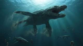
On le croyait roi des océans au Jurassique : une vertèbre percée raconte un combat d’une violence extrême
os dent vertèbre chasse musée Jurassique fossile Ichthyosauria Ichthyosaurus Pliosaurus comportement
Oublié pendant plus d’un siècle dans les tiroirs d’un musée, un fossile refait aujourd’hui surface et raconte une scène de violence extrême dans les océans du Jurassique. Une vertèbre d’ichtyosaure, marquée par l’impact d’une dent brisée, révèle peut-être l’attaque d’un des plus redoutables...
17/06/2026 futura-terre
Description d'un fossile de dinosaure rare du groupe de Nanaimo du Crétacé supérieur
os vertèbre description Canada Colombie Crétacé Crétacé supérieur fossile Dinosauria Ornithomimosauria
Les chercheurs ont décrit une seule vertèbre caudale de dinosaure (coccyx) de l'île Denman (Colombie-Britannique, Canada).  Il a été identifié comme une vertèbre caudale d'ornithomimosaure. Le fossile, censé représenter un os de la partie médiane de la queue, n'est que le deuxième fossile de dinosaure identifié dans le groupe de Nanaimo du Crétacé supérieur. De plus, il
10/06/2026 everythingdinosaur ⚙ Traduction automatique
Ce rapace récemment découvert aurait chassé comme un héron géant
vertèbre prédateur Dinosauria Kank
Un dinosaure ressemblant à un rapace récemment découvert en Patagonie change la façon dont les scientifiques perçoivent les anciens prédateurs. Nommé Kank australis, ce dinosaure vieux de 70 millions d'années semble avoir chassé le poisson un peu comme les hérons modernes, en utilisant un cou long et flexible et des vertèbres spécialisées adaptées pour des mouvements rapides et précis.
29/05/2026 sciencedaily ⚙ Traduction automatique
1 2 3