vertèbre

Partie anatomique

80 image(s) · 11 Actualités

Galerie d'images

FSAC-OB 232, holotype skeleton of Barbaridactylus grandis.
(A) Left humerus in anterior view, (B) right femur in anterior view, (C) right radius and ulna in posterior view, (D) cervical vertebra in ventral view, (E) left scapulocoracoid in medial view, and (F) posterior ramus of the right mandible in medial view.

Abbreviations: co, coracoid; cot, cotyle of mandible; cvr, cervical ribs; dm, dorsal margin of mandible; dpc, deltopectoral crest; gl, glenoid; fh, femoral head; gt, greater trochanter; hh, humeral head; hyp, hypapophysis; olp, olecranon process; pex, postexapophysis; pn, pneumatopore; rad, radius; sca, scapula; scp, supracondylar process of the ectepicondyle; uc, ulnar crest; uln, ulna; vm, ventral margin of mandible.
Taxons Barbaridactylus

FSAC-OB 232, holotype skeleton of Barbaridactylus grandis. (A) Left humerus in anterior view, (B) right femur in anterior view, (C) right radius and ulna in posterior view, (D) cervical vertebra in ventral view, (E) left scapulocoracoid in medial view, and (F) posterior ramus of the right mandible in medial view. Abbreviations: co, coracoid; cot, cotyle of mandible; cvr, cervical ribs; dm, dorsal margin of mandible; dpc, deltopectoral crest; gl, glenoid; fh, femoral head; gt, greater trochanter; hh, humeral head; hyp, hypapophysis; olp, olecranon process; pex, postexapophysis; pn, pneumatopore; rad, radius; sca, scapula; scp, supracondylar process of the ectepicondyle; uc, ulnar crest; uln, ulna; vm, ventral margin of mandible.

crête humérus vertèbre holotype +2
Haliskia peterseni gen. et sp. nov., holotype specimen KK F1426. (A) dorsal vertebra, ribs, gastralium, left scapulocoracoid, left and right manual phalanx IV-2, right manual phalanx IV-3, left femur, left tibia, metatarsals and pedal phalanges, (B) mandible, teeth, ceratobranchials, cervical vertebra, rib, gastralium, right syncarpus, right lateral carpal, right pteroid, metacarpals, manual digits, right manual phalanx IV-1, left manual phalanx IV-4, (C) premaxilla with teeth, (D) isolated tooth, (E) isolated tooth, (F) left lateral carpal, (G) left metacarpal IV. Scale bar = 50 mm.
Taxons Haliskia

Haliskia peterseni gen. et sp. nov., holotype specimen KK F1426. (A) dorsal vertebra, ribs, gastralium, left scapulocoracoid, left and right manual phalanx IV-2, right manual phalanx IV-3, left femur, left tibia, metatarsals and pedal phalanges, (B) mandible, teeth, ceratobranchials, cervical vertebra, rib, gastralium, right syncarpus, right lateral carpal, right pteroid, metacarpals, manual digits, right manual phalanx IV-1, left manual phalanx IV-4, (C) premaxilla with teeth, (D) isolated tooth, (E) isolated tooth, (F) left lateral carpal, (G) left metacarpal IV. Scale bar = 50 mm.

écaille dent vertèbre holotype +2
Entire skeleton of Jidapterus edentus (RCPS-030366CY) preserved in a slab mostly in right lateral view.

Abbreviations: aa, the atlas-axis complex; bo, basioccipital; br, braincase; cve, cervical vertebrae; cv3, the third cervical vertebra; cv8, the eighth cervical vertebra; ds, dentaries; dv, dorsal vertebra; dve, dorsal vertebrae; ep, epiphysis; j, jugal; lc, left carpus; lf, left femur; li, left ilium; lis, left ischium; lm, left manual digits; lp, left pubis; lpe, left pes; lpt, left pteroid; lr, left radius; lsc, left scapula-coracoid; lt, left tibia; lta, left tarsus; lu, left ulna; lwmc, left wing metacarpal; lwp1–4; left wing phalanges 1–4; p, pterygoid; pp, postacetabular process of the right ilium; q, quadrate; rc, right carpus; rh, right humerus; rf, right femur; rfi, right fibula; ribs, ribs; ris, right ischium; rm, right manual digits; rmcI, right metacarpal I; rp, right pubis; rpe, right pes; rpt, right pteroid; rr, right radius; rsc, right scapula-coracoid; rt, right tibia; ru, right ulna; rwmc, right wing metacarpal; rwp1–4, right wing phalanges 1–4; rostrum, rostrum; st, sternum; sv, sacral vertebra; sve, sacral vertebrae. Gray shaded areas bound by solid lines indicate preserved bones, dashed lines indicate bones with margins discerned by impression, and lack of lines indicate missing bone or displacement along cracks in the slab.
Taxons Jidapterus

Entire skeleton of Jidapterus edentus (RCPS-030366CY) preserved in a slab mostly in right lateral view. Abbreviations: aa, the atlas-axis complex; bo, basioccipital; br, braincase; cve, cervical vertebrae; cv3, the third cervical vertebra; cv8, the eighth cervical vertebra; ds, dentaries; dv, dorsal vertebra; dve, dorsal vertebrae; ep, epiphysis; j, jugal; lc, left carpus; lf, left femur; li, left ilium; lis, left ischium; lm, left manual digits; lp, left pubis; lpe, left pes; lpt, left pteroid; lr, left radius; lsc, left scapula-coracoid; lt, left tibia; lta, left tarsus; lu, left ulna; lwmc, left wing metacarpal; lwp1–4; left wing phalanges 1–4; p, pterygoid; pp, postacetabular process of the right ilium; q, quadrate; rc, right carpus; rh, right humerus; rf, right femur; rfi, right fibula; ribs, ribs; ris, right ischium; rm, right manual digits; rmcI, right metacarpal I; rp, right pubis; rpe, right pes; rpt, right pteroid; rr, right radius; rsc, right scapula-coracoid; rt, right tibia; ru, right ulna; rwmc, right wing metacarpal; rwp1–4, right wing phalanges 1–4; rostrum, rostrum; st, sternum; sv, sacral vertebra; sve, sacral vertebrae. Gray shaded areas bound by solid lines indicate preserved bones, dashed lines indicate bones with margins discerned by impression, and lack of lines indicate missing bone or displacement along cracks in the slab.

humérus vertèbre Chaoyangopteridae Chaoyangopterinae +2
Entire skeleton of Jidapterus edentus (RCPS-030366CY) preserved in a slab mostly in right lateral view.

Abbreviations: aa, the atlas-axis complex; bo, basioccipital; br, braincase; cve, cervical vertebrae; cv3, the third cervical vertebra; cv8, the eighth cervical vertebra; ds, dentaries; dv, dorsal vertebra; dve, dorsal vertebrae; ep, epiphysis; j, jugal; lc, left carpus; lf, left femur; li, left ilium; lis, left ischium; lm, left manual digits; lp, left pubis; lpe, left pes; lpt, left pteroid; lr, left radius; lsc, left scapula-coracoid; lt, left tibia; lta, left tarsus; lu, left ulna; lwmc, left wing metacarpal; lwp1–4; left wing phalanges 1–4; p, pterygoid; pp, postacetabular process of the right ilium; q, quadrate; rc, right carpus; rh, right humerus; rf, right femur; rfi, right fibula; ribs, ribs; ris, right ischium; rm, right manual digits; rmcI, right metacarpal I; rp, right pubis; rpe, right pes; rpt, right pteroid; rr, right radius; rsc, right scapula-coracoid; rt, right tibia; ru, right ulna; rwmc, right wing metacarpal; rwp1–4, right wing phalanges 1–4; rostrum, rostrum; st, sternum; sv, sacral vertebra; sve, sacral vertebrae. Gray shaded areas bound by solid lines indicate preserved bones, dashed lines indicate bones with margins discerned by impression, and lack of lines indicate missing bone or displacement along cracks in the slab.
Taxons Chaoyangopterinae

Entire skeleton of Jidapterus edentus (RCPS-030366CY) preserved in a slab mostly in right lateral view. Abbreviations: aa, the atlas-axis complex; bo, basioccipital; br, braincase; cve, cervical vertebrae; cv3, the third cervical vertebra; cv8, the eighth cervical vertebra; ds, dentaries; dv, dorsal vertebra; dve, dorsal vertebrae; ep, epiphysis; j, jugal; lc, left carpus; lf, left femur; li, left ilium; lis, left ischium; lm, left manual digits; lp, left pubis; lpe, left pes; lpt, left pteroid; lr, left radius; lsc, left scapula-coracoid; lt, left tibia; lta, left tarsus; lu, left ulna; lwmc, left wing metacarpal; lwp1–4; left wing phalanges 1–4; p, pterygoid; pp, postacetabular process of the right ilium; q, quadrate; rc, right carpus; rh, right humerus; rf, right femur; rfi, right fibula; ribs, ribs; ris, right ischium; rm, right manual digits; rmcI, right metacarpal I; rp, right pubis; rpe, right pes; rpt, right pteroid; rr, right radius; rsc, right scapula-coracoid; rt, right tibia; ru, right ulna; rwmc, right wing metacarpal; rwp1–4, right wing phalanges 1–4; rostrum, rostrum; st, sternum; sv, sacral vertebra; sve, sacral vertebrae. Gray shaded areas bound by solid lines indicate preserved bones, dashed lines indicate bones with margins discerned by impression, and lack of lines indicate missing bone or displacement along cracks in the slab.

humérus vertèbre Chaoyangopteridae Chaoyangopterinae +2
Nemicolopterus crypticus holotype (IVPP V-14377).

(A) Skeleton overview, and (B) schematic drawing. (C) Skull (right lateral view), and (D) schematic drawing. Abbreviations: cdv, caudal vertebrae; co, coracoid; cv, cervical vertebra; d1–d4, digits 1–4; f, frontal; fe, femur; h, humerus; hy, hyoid; il, illium; is, ischium; j, jugal; l, left; la, lacrimal; mand, mandible; mc, metacarpal; mt, metatarsal; naof, nasoantorbital fenestra; or, orbit; pa, parietal; pm, premaxilla; ph, phalanx; ti, tibia; r, right; sca, scapula. Scale bars: A–B, 100 mm; C–D, 5 mm.
Taxons Nemicolopterus

Nemicolopterus crypticus holotype (IVPP V-14377). (A) Skeleton overview, and (B) schematic drawing. (C) Skull (right lateral view), and (D) schematic drawing. Abbreviations: cdv, caudal vertebrae; co, coracoid; cv, cervical vertebra; d1–d4, digits 1–4; f, frontal; fe, femur; h, humerus; hy, hyoid; il, illium; is, ischium; j, jugal; l, left; la, lacrimal; mand, mandible; mc, metacarpal; mt, metatarsal; naof, nasoantorbital fenestra; or, orbit; pa, parietal; pm, premaxilla; ph, phalanx; ti, tibia; r, right; sca, scapula. Scale bars: A–B, 100 mm; C–D, 5 mm.

humérus écaille vertèbre dessin +4
Ceratosaurian theropod Camarillasaurus cirugedae gen. et sp. nov. from the Camarillas Formation of Camarillas, Soria Province, Spain, presacral vertebra, MPG-KPC9,  in anterior (A), left lateral (B), and posterior (C) views; possible neural spine tips, MPG1116, 32, 33 in ?posterior (D) and transverse (E) views; and presacral rib MPG-KPC7, in ventral view (F) and detail of its proximal end (G). Scale bars are 10 mm (A-C, G), 20 mm (D-E) and 100 mm (F).

Ceratosaurian theropod Camarillasaurus cirugedae gen. et sp. nov. from the Camarillas Formation of Camarillas, Soria Province, Spain, presacral vertebra, MPG-KPC9, in anterior (A), left lateral (B), and posterior (C) views; possible neural spine tips, MPG1116, 32, 33 in ?posterior (D) and transverse (E) views; and presacral rib MPG-KPC7, in ventral view (F) and detail of its proximal end (G). Scale bars are 10 mm (A-C, G), 20 mm (D-E) and 100 mm (F).

écaille vertèbre Espagne Camarillas +3
Skeletal reconstruction and postcranial anatomy of Asfaltovenator vialidadi, MPEF PV 3440. Centre: body outline with preserved elements indicated. (A) articulated cervical vertebrae three to five. (B) cervical vertebra 7. (C) articulated dorsal vertebrae four to seven (better preserved right side, reversed). (D) right humerus in anterior view. (E) right radius and ulna, medial view. (F) right manus, metacarpus in dorsal and digits in lateral view. (G) articulated proximal ends of right tibia and fibula in lateral and proximal views. Abbreviations: ag, anterior groove; cc, cnemial crest; cr, cervical rib; dc, distal carpal; di, diapophysis; dpc, deltpectoral crest; ec, ectepicondyle; ent, entepicondyle; epi, epipophysis; fi, fibula; fic, fibular condyle; hy, hyposphene; im, intermedium; it, internal tuberosity; lr, lateral ridge; mc, metacarpal; ns, neural spine; ol, olecranon; pa, parapophysis; pl, pleurocoel; poz, postzygapophysis; ppdl, paradiapophyseal lamina; prz, prezygapophysis; ra, radial. Scale bars are 100 cm (skeletal reconstruction), 5 cm (A–C) and 10 cm (D–G).
Taxons Asfaltovenator

Skeletal reconstruction and postcranial anatomy of Asfaltovenator vialidadi, MPEF PV 3440. Centre: body outline with preserved elements indicated. (A) articulated cervical vertebrae three to five. (B) cervical vertebra 7. (C) articulated dorsal vertebrae four to seven (better preserved right side, reversed). (D) right humerus in anterior view. (E) right radius and ulna, medial view. (F) right manus, metacarpus in dorsal and digits in lateral view. (G) articulated proximal ends of right tibia and fibula in lateral and proximal views. Abbreviations: ag, anterior groove; cc, cnemial crest; cr, cervical rib; dc, distal carpal; di, diapophysis; dpc, deltpectoral crest; ec, ectepicondyle; ent, entepicondyle; epi, epipophysis; fi, fibula; fic, fibular condyle; hy, hyposphene; im, intermedium; it, internal tuberosity; lr, lateral ridge; mc, metacarpal; ns, neural spine; ol, olecranon; pa, parapophysis; pl, pleurocoel; poz, postzygapophysis; ppdl, paradiapophyseal lamina; prz, prezygapophysis; ra, radial. Scale bars are 100 cm (skeletal reconstruction), 5 cm (A–C) and 10 cm (D–G).

crête humérus écaille vertèbre +3
Becklespinax altispinax holotype BMNH R1828 (originally assigned to Megalosaurus bucklandii). Owen's description follows:
Three anterior dorsal vertebræ : p, parapophysis, or lower transverse process: t, accessory tubercle contributing some attachment to the head of the rib : d, diapophyses, or upper transverse process, fractured, which gave attachment to the tubercle of the rib: b, oblique buttress extending from the parapophysis to the diapophysis, and contributing to the support of the neural platform : z, the prozygapophysis, z', the zygapophysis, forming the ends of the neural platform and articulating the neural arches of the vertebræ with each other, ns, the neural spine of the foremost of these vertebras, ns', the neural spine of the second vertebra; it expands at its extremity, overhangs the anterior shorter spine, and developes a strong bony plate from its back part which fixes it to n", the similarly developed and modified spine of the third vertebra.
The extraordinary size and strength of the spines of these anterior dorsal vertebræ, indicate the great force with which the head and jaws of the Megalosaurus must have been used.

From the Wealden, near Battle. In the Museum of Samuel H. Beckles, Esq., F.G.S.
Taxons Becklespinax

Becklespinax altispinax holotype BMNH R1828 (originally assigned to Megalosaurus bucklandii). Owen's description follows: Three anterior dorsal vertebræ : p, parapophysis, or lower transverse process: t, accessory tubercle contributing some attachment to the head of the rib : d, diapophyses, or upper transverse process, fractured, which gave attachment to the tubercle of the rib: b, oblique buttress extending from the parapophysis to the diapophysis, and contributing to the support of the neural platform : z, the prozygapophysis, z', the zygapophysis, forming the ends of the neural platform and articulating the neural arches of the vertebræ with each other, ns, the neural spine of the foremost of these vertebras, ns', the neural spine of the second vertebra; it expands at its extremity, overhangs the anterior shorter spine, and developes a strong bony plate from its back part which fixes it to n", the similarly developed and modified spine of the third vertebra. The extraordinary size and strength of the spines of these anterior dorsal vertebræ, indicate the great force with which the head and jaws of the Megalosaurus must have been used. From the Wealden, near Battle. In the Museum of Samuel H. Beckles, Esq., F.G.S.

vertèbre description Foremost holotype +3
Becklespinax altispinax holotype BMNH R1828 (originally assigned to Megalosaurus bucklandii). Owen's description follows:
Three anterior dorsal vertebræ : p, parapophysis, or lower transverse process: t, accessory tubercle contributing some attachment to the head of the rib : d, diapophyses, or upper transverse process, fractured, which gave attachment to the tubercle of the rib: b, oblique buttress extending from the parapophysis to the diapophysis, and contributing to the support of the neural platform : z, the prozygapophysis, z', the zygapophysis, forming the ends of the neural platform and articulating the neural arches of the vertebræ with each other, ns, the neural spine of the foremost of these vertebras, ns', the neural spine of the second vertebra; it expands at its extremity, overhangs the anterior shorter spine, and developes a strong bony plate from its back part which fixes it to n", the similarly developed and modified spine of the third vertebra.
The extraordinary size and strength of the spines of these anterior dorsal vertebræ, indicate the great force with which the head and jaws of the Megalosaurus must have been used.

From the Wealden, near Battle. In the Museum of Samuel H. Beckles, Esq., F.G.S.
Taxons Altispinax

Becklespinax altispinax holotype BMNH R1828 (originally assigned to Megalosaurus bucklandii). Owen's description follows: Three anterior dorsal vertebræ : p, parapophysis, or lower transverse process: t, accessory tubercle contributing some attachment to the head of the rib : d, diapophyses, or upper transverse process, fractured, which gave attachment to the tubercle of the rib: b, oblique buttress extending from the parapophysis to the diapophysis, and contributing to the support of the neural platform : z, the prozygapophysis, z', the zygapophysis, forming the ends of the neural platform and articulating the neural arches of the vertebræ with each other, ns, the neural spine of the foremost of these vertebras, ns', the neural spine of the second vertebra; it expands at its extremity, overhangs the anterior shorter spine, and developes a strong bony plate from its back part which fixes it to n", the similarly developed and modified spine of the third vertebra. The extraordinary size and strength of the spines of these anterior dorsal vertebræ, indicate the great force with which the head and jaws of the Megalosaurus must have been used. From the Wealden, near Battle. In the Museum of Samuel H. Beckles, Esq., F.G.S.

vertèbre description Foremost holotype +3
Caihong juji holotype specimen (PMoL-B00175). Photographs of the slab (a) and counter slab (b) and line drawing (c) of the specimen based on both slabs. Photograph (d) and line drawing (e) of a composite of the rostrum of the skull and mandible exposed on the counter slab and the post-rostrum cranium exposed on the slab. Arrows indicate lacrimal crests. Question mark indicates uncertain identification. Scale bars: 10 cm a–c, 1 cm d and e. aof antorbital fenestra, cav caudal vertebra, cev cervical vertebra, dr dorsal rib, dv dorsal vertebra, ect ectopterygoid, emf external mandibular fenestra, en external naris, f feather, fu furcula, ga gastralia, hy hyoid, il ilium, is ischium, la left angular, lar left articular, lc left coracoid, lcr lacrimal crest, ld left dentary, lf left, frontal, lfe left femur, lh left humerus, lj left jugal, ll left lacrimal, lma left maxilla, lm left manus, ln left nasal, lp left pes, lpa left palatine, lpo left postorbital, lq left quadrate, lqj left quadratojugal, lr left radius, ls left scapula, lsp left splenial, lsa left surangular, lsq left squamosal, lt left tibiotarsus, lu left ulna, ma mandible, mf maxillary fenestra, o orbit, p parietal, pm premaxilla, pt pterygoid, pu pubis, rar right articular, rc right coracoid, rd right dentary, rfe right femur, rh right humerus, rm right manus, rp right pes, rpra right prearticular, rq right quadrate, rr right radius, rs right scapula, rt right tibiotarsus, ru right ulna, scl sclerotic bones, sk skull, sy synsacrum
Taxons Caihong

Caihong juji holotype specimen (PMoL-B00175). Photographs of the slab (a) and counter slab (b) and line drawing (c) of the specimen based on both slabs. Photograph (d) and line drawing (e) of a composite of the rostrum of the skull and mandible exposed on the counter slab and the post-rostrum cranium exposed on the slab. Arrows indicate lacrimal crests. Question mark indicates uncertain identification. Scale bars: 10 cm a–c, 1 cm d and e. aof antorbital fenestra, cav caudal vertebra, cev cervical vertebra, dr dorsal rib, dv dorsal vertebra, ect ectopterygoid, emf external mandibular fenestra, en external naris, f feather, fu furcula, ga gastralia, hy hyoid, il ilium, is ischium, la left angular, lar left articular, lc left coracoid, lcr lacrimal crest, ld left dentary, lf left, frontal, lfe left femur, lh left humerus, lj left jugal, ll left lacrimal, lma left maxilla, lm left manus, ln left nasal, lp left pes, lpa left palatine, lpo left postorbital, lq left quadrate, lqj left quadratojugal, lr left radius, ls left scapula, lsp left splenial, lsa left surangular, lsq left squamosal, lt left tibiotarsus, lu left ulna, ma mandible, mf maxillary fenestra, o orbit, p parietal, pm premaxilla, pt pterygoid, pu pubis, rar right articular, rc right coracoid, rd right dentary, rfe right femur, rh right humerus, rm right manus, rp right pes, rpra right prearticular, rq right quadrate, rr right radius, rs right scapula, rt right tibiotarsus, ru right ulna, scl sclerotic bones, sk skull, sy synsacrum

crête plume humérus écaille +7
Skeletal reconstruction of Dineobellator notohesperus gen. et sp. nov., SMP VP-2430, with known elements colored in white. Figured bones are as follows: fused distal caudal vertebra (A); middle caudal vertebra (B); caudal vertebra 1 (C); right femur (D); rib (E); right basipterygoid (F); left lacrimal (reversed) (G); right jugal (H); right humerus (I); right ulna (J); right metacarpal III (K); right manual ungual II (L); right metatarsal II (M); right metatarsal III. (N) Individual scale bars, 2 cm. Skeletal drawing based off work of Scott Hartman.
Taxons Dineobellator

Skeletal reconstruction of Dineobellator notohesperus gen. et sp. nov., SMP VP-2430, with known elements colored in white. Figured bones are as follows: fused distal caudal vertebra (A); middle caudal vertebra (B); caudal vertebra 1 (C); right femur (D); rib (E); right basipterygoid (F); left lacrimal (reversed) (G); right jugal (H); right humerus (I); right ulna (J); right metacarpal III (K); right manual ungual II (L); right metatarsal II (M); right metatarsal III. (N) Individual scale bars, 2 cm. Skeletal drawing based off work of Scott Hartman.

os humérus écaille vertèbre +2
(A) Skeletal reconstruction in left lateral view (missing and damaged portions of the bones in gray). (B) Left ilium in lateral view. (C) Proximal caudal vertebrae in left lateral view with close-up of the infraprezygapophyses. (D) Chevron in cranial view. (E-F) Right scapula in dorsal (E) and lateral (F) views. (G) Last sacral and the two proximalmost caudals in left lateral view. (H) Right pubis in medial view. (I) Right ischium in lateral view. (J) Right femur in distal view. (K) Left metatarsus and distal tarsals in proximal view. (L) Right femur in cranial view. (M-N) Left metatarsus in lateral (M) and dorsal (N) views. Abbreviations: acr, acromion process; ant, antitrochanter; ch, chevron; cv, caudal vertebra(e); diprf, dorsal infraprezygapophyseal fossa; dt, distal tarsal(s); fct, cranial trochanter of femur; fh, femoral head; gl, glenoid fossa; idf, infradiapophyseal fossa; lc, lateral condyle; mc, medial condyle; mep, medial epicondyle; miprf, middle infraprezygapophyseal fossa; mt II, metatarsal II; mt IV, metatarsal IV; mt V, metatarsal V; ns, neural spine; obp, obturator process; pra, preacetabular process; pup, pubic peduncle; sprf, supraprezygapophyseal fossa; sv, sacral vertebra; tfc, tibiofibular crest; tp, transverse process; viprf, ventral infraprezygapophyseal fossa. Scale bars equal 10 cm in (A); 1 cm in (B-N).
https://doi.org/10.1371/journal.pone.0210867.g004
Taxons Gobiraptor

(A) Skeletal reconstruction in left lateral view (missing and damaged portions of the bones in gray). (B) Left ilium in lateral view. (C) Proximal caudal vertebrae in left lateral view with close-up of the infraprezygapophyses. (D) Chevron in cranial view. (E-F) Right scapula in dorsal (E) and lateral (F) views. (G) Last sacral and the two proximalmost caudals in left lateral view. (H) Right pubis in medial view. (I) Right ischium in lateral view. (J) Right femur in distal view. (K) Left metatarsus and distal tarsals in proximal view. (L) Right femur in cranial view. (M-N) Left metatarsus in lateral (M) and dorsal (N) views. Abbreviations: acr, acromion process; ant, antitrochanter; ch, chevron; cv, caudal vertebra(e); diprf, dorsal infraprezygapophyseal fossa; dt, distal tarsal(s); fct, cranial trochanter of femur; fh, femoral head; gl, glenoid fossa; idf, infradiapophyseal fossa; lc, lateral condyle; mc, medial condyle; mep, medial epicondyle; miprf, middle infraprezygapophyseal fossa; mt II, metatarsal II; mt IV, metatarsal IV; mt V, metatarsal V; ns, neural spine; obp, obturator process; pra, preacetabular process; pup, pubic peduncle; sprf, supraprezygapophyseal fossa; sv, sacral vertebra; tfc, tibiofibular crest; tp, transverse process; viprf, ventral infraprezygapophyseal fossa. Scale bars equal 10 cm in (A); 1 cm in (B-N). https://doi.org/10.1371/journal.pone.0210867.g004

os vertèbre Gobiraptor
A photograph of the skeleton of Jianchangosaurus yixianensis gen. et sp. nov. (41HIII-0308A) (A) and line drawing (B).

Abbreviations: at, atlas; ax, axis; cav, caudal vertebra; cv, cervical vertebra; co, coracoid; dc, distal carpal; dv, dorsal vertebra; l., left; f, femur; fi, fibula; fur, furcula; hu, humerus; il, ilium; is, ischium; pu, pubis; ra, radius; sc, scapula; ti, tibia; ul, ulna; mc, metacarpal; mt, metatarsal; mxf, maxillary fenestra; pat, proatlas; r., right; sr, sacral rib; sv, sacral vertebra; ung, ungual. All elements of the skeleton are preserved except the distal half of the caudal vertebrae. Dashed lines of metatarsals indicate areas that have been reconstructed. The middle portion of the neck, from the fourth to ninth cervical vertebrae, and the pedal phalanges have been repositioned. The rest of elements of this specimen are in the original position.
Taxons Jianchangosaurus

A photograph of the skeleton of Jianchangosaurus yixianensis gen. et sp. nov. (41HIII-0308A) (A) and line drawing (B). Abbreviations: at, atlas; ax, axis; cav, caudal vertebra; cv, cervical vertebra; co, coracoid; dc, distal carpal; dv, dorsal vertebra; l., left; f, femur; fi, fibula; fur, furcula; hu, humerus; il, ilium; is, ischium; pu, pubis; ra, radius; sc, scapula; ti, tibia; ul, ulna; mc, metacarpal; mt, metatarsal; mxf, maxillary fenestra; pat, proatlas; r., right; sr, sacral rib; sv, sacral vertebra; ung, ungual. All elements of the skeleton are preserved except the distal half of the caudal vertebrae. Dashed lines of metatarsals indicate areas that have been reconstructed. The middle portion of the neck, from the fourth to ninth cervical vertebrae, and the pedal phalanges have been repositioned. The rest of elements of this specimen are in the original position.

humérus vertèbre dessin spécimen +2
Alvarezsauridae gen. et sp. indet., posterior caudal vertebrae. Dzharakuduk, Uzbekistan; Bissekty Formation, Upper Cretaceous (Turonian). a-e, ZIN PH 2441/16, two vertebrae preserved in articulation, in anterior (a), dorsal (b), posterior (c), lateral (d), and ventral (e) views. f-j, ZIN PH 2442/16, vertebra missing ventral part of the centrum, in dorsal (f), lateral (g), ventral (h), anterior (i), and posterior (j).
Taxons Dzharaonyx

Alvarezsauridae gen. et sp. indet., posterior caudal vertebrae. Dzharakuduk, Uzbekistan; Bissekty Formation, Upper Cretaceous (Turonian). a-e, ZIN PH 2441/16, two vertebrae preserved in articulation, in anterior (a), dorsal (b), posterior (c), lateral (d), and ventral (e) views. f-j, ZIN PH 2442/16, vertebra missing ventral part of the centrum, in dorsal (f), lateral (g), ventral (h), anterior (i), and posterior (j).

vertèbre Ouzbékistan Bissekty Crétacé +4
Cervical vertebra of Calamosaurus foxi
Taxons Calamosaurus

Cervical vertebra of Calamosaurus foxi

vertèbre Calamosaurus
Tetanuran theropod Cruxicheiros newmanorum gen. et sp. nov. axial vertebrae (WARMS 15771) from the Chipping Norton Limestone Formation,
Bathonian of the United Kingdom. A. Posterior cervical or anterior dorsal vertebra in posterior view. B. Partial middle−posterior dorsal vertebra in right lat−
eral view showing a sagittal cross−section (B1) and in dorsal view (B2). C. Middle−distal caudal vertebra in left lateral (C1) and posterior (C2) views. D. Mid−
dle−posterior dorsal neural arch in anterior (D1), right lateral (D2, D3), and posterior (D4) views. Photographs (A, B1, B2, C1, C2, D1, D2, D4) and line drawing

(D3). Crossed−hatching indicates matrix and grey tone indicates broken bone.
Taxons Cruxicheiros

Tetanuran theropod Cruxicheiros newmanorum gen. et sp. nov. axial vertebrae (WARMS 15771) from the Chipping Norton Limestone Formation, Bathonian of the United Kingdom. A. Posterior cervical or anterior dorsal vertebra in posterior view. B. Partial middle−posterior dorsal vertebra in right lat− eral view showing a sagittal cross−section (B1) and in dorsal view (B2). C. Middle−distal caudal vertebra in left lateral (C1) and posterior (C2) views. D. Mid− dle−posterior dorsal neural arch in anterior (D1), right lateral (D2, D3), and posterior (D4) views. Photographs (A, B1, B2, C1, C2, D1, D2, D4) and line drawing (D3). Crossed−hatching indicates matrix and grey tone indicates broken bone.

os vertèbre dessin Royaume-Uni +4
1 2 3 4 5

Actualités

Des scientifiques décrivent officiellement le premier fossile de dinosaure découvert en Antarctique
vertèbre Antarctique Crétacé Crétacé supérieur fossile Dinosauria Titanosauria découverte
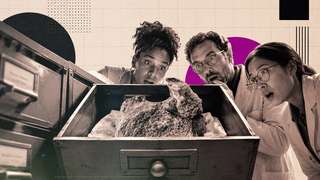
Les scientifiques ont officiellement décrit le premier fossile de dinosaure découvert en Antarctique. La découverte remarquable est une seule vertèbre provenant d’un titanosaure du Crétacé supérieur. Bien que le fossile ait été collecté il y a plus de quarante ans, les chercheurs n’ont confirmé son identité que récemment.  Le premier fossile de dinosaure découvert en Antarctique est décrit dans la revue Acta Palaeontologica Polonica.
29/06/2026 everythingdinosaur ⚙ Traduction automatique
On le croyait roi des océans au Jurassique : une vertèbre percée raconte un combat d’une violence extrême
os dent vertèbre chasse musée Jurassique fossile Ichthyosauria Ichthyosaurus Pliosaurus comportement
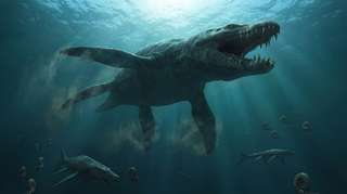
Oublié pendant plus d’un siècle dans les tiroirs d’un musée, un fossile refait aujourd’hui surface et raconte une scène de violence extrême dans les océans du Jurassique. Une vertèbre d’ichtyosaure, marquée par l’impact d’une dent brisée, révèle peut-être l’attaque d’un des plus redoutables...
17/06/2026 futura-terre
Description d'un fossile de dinosaure rare du groupe de Nanaimo du Crétacé supérieur
os vertèbre description Canada Colombie Crétacé Crétacé supérieur fossile Dinosauria Ornithomimosauria
Les chercheurs ont décrit une seule vertèbre caudale de dinosaure (coccyx) de l'île Denman (Colombie-Britannique, Canada).  Il a été identifié comme une vertèbre caudale d'ornithomimosaure. Le fossile, censé représenter un os de la partie médiane de la queue, n'est que le deuxième fossile de dinosaure identifié dans le groupe de Nanaimo du Crétacé supérieur. De plus, il
10/06/2026 everythingdinosaur ⚙ Traduction automatique
Ce rapace récemment découvert aurait chassé comme un héron géant
vertèbre prédateur Dinosauria Kank
Un dinosaure ressemblant à un rapace récemment découvert en Patagonie change la façon dont les scientifiques perçoivent les anciens prédateurs. Nommé Kank australis, ce dinosaure vieux de 70 millions d'années semble avoir chassé le poisson un peu comme les hérons modernes, en utilisant un cou long et flexible et des vertèbres spécialisées adaptées pour des mouvements rapides et précis.
29/05/2026 sciencedaily ⚙ Traduction automatique
Rare Ostrich-Like Dinosaur Fossil Found on Canadian Island
Un rare fossile de dinosaure ressemblant à une autruche découvert sur une île canadienne
vertèbre Canada Colombie fossile Dinosauria Ornithomimosauria oiseau
Des paléontologues canadiens affirment avoir découvert une vertèbre caudale de dinosaure dans des roches marines vieilles de 75 à 80 millions d'années sur une petite île au large de la Colombie-Britannique, fournissant ainsi la preuve la plus claire à ce jour que des ornithomimosaures ressemblant à des oiseaux parcouraient autrefois l'ancienne côte du Pacifique de l'Amérique du Nord. L'article Un fossile rare de dinosaure ressemblant à une autruche trouvé sur une île canadienne est apparu en premier sur Sci.News : Breaking Science News.
25/05/2026 sci-news ⚙ Traduction automatique
1 2 3