←

vertèbre

Partie anatomique

70 image(s) · 12 Actualités

Galerie d'images

The holotype of Douzhanopterus zhengi gen. et sp. nov.

(a) Part of the holotype; (b) close up of the tail, green arrows indicating the anterior and posterior ends of each caudal vertebra; (c) close up of the right foot. Scale bars are 50 mm, 10 mm, and 10 mm in (a,b and c), respectively.
Taxons Douzhanopterus

The holotype of Douzhanopterus zhengi gen. et sp. nov. (a) Part of the holotype; (b) close up of the tail, green arrows indicating the anterior and posterior ends of each caudal vertebra; (c) close up of the right foot. Scale bars are 50 mm, 10 mm, and 10 mm in (a,b and c), respectively.

écaille vertèbre holotype Douzhanopterus
Haliskia peterseni gen. et sp. nov., holotype specimen KK F1426. (A) dorsal vertebra, ribs, gastralium, left scapulocoracoid, left and right manual phalanx IV-2, right manual phalanx IV-3, left femur, left tibia, metatarsals and pedal phalanges, (B) mandible, teeth, ceratobranchials, cervical vertebra, rib, gastralium, right syncarpus, right lateral carpal, right pteroid, metacarpals, manual digits, right manual phalanx IV-1, left manual phalanx IV-4, (C) premaxilla with teeth, (D) isolated tooth, (E) isolated tooth, (F) left lateral carpal, (G) left metacarpal IV. Scale bar = 50 mm.
Taxons Haliskia

Haliskia peterseni gen. et sp. nov., holotype specimen KK F1426. (A) dorsal vertebra, ribs, gastralium, left scapulocoracoid, left and right manual phalanx IV-2, right manual phalanx IV-3, left femur, left tibia, metatarsals and pedal phalanges, (B) mandible, teeth, ceratobranchials, cervical vertebra, rib, gastralium, right syncarpus, right lateral carpal, right pteroid, metacarpals, manual digits, right manual phalanx IV-1, left manual phalanx IV-4, (C) premaxilla with teeth, (D) isolated tooth, (E) isolated tooth, (F) left lateral carpal, (G) left metacarpal IV. Scale bar = 50 mm.

écaille dent vertèbre holotype +2
Skeletal reconstruction and postcranial anatomy of Asfaltovenator vialidadi, MPEF PV 3440. Centre: body outline with preserved elements indicated. (A) articulated cervical vertebrae three to five. (B) cervical vertebra 7. (C) articulated dorsal vertebrae four to seven (better preserved right side, reversed). (D) right humerus in anterior view. (E) right radius and ulna, medial view. (F) right manus, metacarpus in dorsal and digits in lateral view. (G) articulated proximal ends of right tibia and fibula in lateral and proximal views. Abbreviations: ag, anterior groove; cc, cnemial crest; cr, cervical rib; dc, distal carpal; di, diapophysis; dpc, deltpectoral crest; ec, ectepicondyle; ent, entepicondyle; epi, epipophysis; fi, fibula; fic, fibular condyle; hy, hyposphene; im, intermedium; it, internal tuberosity; lr, lateral ridge; mc, metacarpal; ns, neural spine; ol, olecranon; pa, parapophysis; pl, pleurocoel; poz, postzygapophysis; ppdl, paradiapophyseal lamina; prz, prezygapophysis; ra, radial. Scale bars are 100 cm (skeletal reconstruction), 5 cm (A–C) and 10 cm (D–G).
Taxons Asfaltovenator

Skeletal reconstruction and postcranial anatomy of Asfaltovenator vialidadi, MPEF PV 3440. Centre: body outline with preserved elements indicated. (A) articulated cervical vertebrae three to five. (B) cervical vertebra 7. (C) articulated dorsal vertebrae four to seven (better preserved right side, reversed). (D) right humerus in anterior view. (E) right radius and ulna, medial view. (F) right manus, metacarpus in dorsal and digits in lateral view. (G) articulated proximal ends of right tibia and fibula in lateral and proximal views. Abbreviations: ag, anterior groove; cc, cnemial crest; cr, cervical rib; dc, distal carpal; di, diapophysis; dpc, deltpectoral crest; ec, ectepicondyle; ent, entepicondyle; epi, epipophysis; fi, fibula; fic, fibular condyle; hy, hyposphene; im, intermedium; it, internal tuberosity; lr, lateral ridge; mc, metacarpal; ns, neural spine; ol, olecranon; pa, parapophysis; pl, pleurocoel; poz, postzygapophysis; ppdl, paradiapophyseal lamina; prz, prezygapophysis; ra, radial. Scale bars are 100 cm (skeletal reconstruction), 5 cm (A–C) and 10 cm (D–G).

crête humérus écaille vertèbre +3
Atsinganosaurus velauciensis (VBN 93.01), late Campanian, Velaux-La Bastide Neuve, Bouches-de-Rhône, southern France. Posterior dorsal vertebra, right lateral view. Scale bar equals 10 cm.
Taxons Atsinganosaurus

Atsinganosaurus velauciensis (VBN 93.01), late Campanian, Velaux-La Bastide Neuve, Bouches-de-Rhône, southern France. Posterior dorsal vertebra, right lateral view. Scale bar equals 10 cm.

écaille vertèbre France Campanien +1
Tetanuran theropod Cruxicheiros newmanorum gen. et sp. nov. axial vertebrae (WARMS 15771) from the Chipping Norton Limestone Formation,
Bathonian of the United Kingdom. A. Posterior cervical or anterior dorsal vertebra in posterior view. B. Partial middle−posterior dorsal vertebra in right lat−
eral view showing a sagittal cross−section (B1) and in dorsal view (B2). C. Middle−distal caudal vertebra in left lateral (C1) and posterior (C2) views. D. Mid−
dle−posterior dorsal neural arch in anterior (D1), right lateral (D2, D3), and posterior (D4) views. Photographs (A, B1, B2, C1, C2, D1, D2, D4) and line drawing

(D3). Crossed−hatching indicates matrix and grey tone indicates broken bone.
Taxons Cruxicheiros

Tetanuran theropod Cruxicheiros newmanorum gen. et sp. nov. axial vertebrae (WARMS 15771) from the Chipping Norton Limestone Formation, Bathonian of the United Kingdom. A. Posterior cervical or anterior dorsal vertebra in posterior view. B. Partial middle−posterior dorsal vertebra in right lat− eral view showing a sagittal cross−section (B1) and in dorsal view (B2). C. Middle−distal caudal vertebra in left lateral (C1) and posterior (C2) views. D. Mid− dle−posterior dorsal neural arch in anterior (D1), right lateral (D2, D3), and posterior (D4) views. Photographs (A, B1, B2, C1, C2, D1, D2, D4) and line drawing (D3). Crossed−hatching indicates matrix and grey tone indicates broken bone.

os vertèbre dessin Royaume-Uni +4
Holotype (HMN MB.R.2455, Cervical vertebra) of Australodocus.
Taxons Qijianglong

Holotype (HMN MB.R.2455, Cervical vertebra) of Australodocus.

vertèbre holotype Australodocus Qijianglong
Holotype (HMN MB.R.2455, Cervical vertebra) of Australodocus.
Taxons Australodocus

Holotype (HMN MB.R.2455, Cervical vertebra) of Australodocus.

vertèbre holotype Australodocus Qijianglong
Figure 1. Holotype NHMUK PV R 37591 pelvis and vertebrae of P. milnerae gen. et sp. nov. in (a) left lateral view and (b) right lateral view. atr, antitrochanter; bf, brevis fossa; bfr, brevis fossa rim; bs, brevis shelf; dv, dorsal vertebra; iss, ischial shaft; nc, neural canal; no, notch; obf, obturator foramen; poap, postacetabular process; prap, preacetabular process; puf, pubic fenestra; pus, pubic shaft; ras, rib attachment scar; ri, rim; sac, supra-acetabular crest; sv, sacral vertebra.
Taxons Pendraig

Figure 1. Holotype NHMUK PV R 37591 pelvis and vertebrae of P. milnerae gen. et sp. nov. in (a) left lateral view and (b) right lateral view. atr, antitrochanter; bf, brevis fossa; bfr, brevis fossa rim; bs, brevis shelf; dv, dorsal vertebra; iss, ischial shaft; nc, neural canal; no, notch; obf, obturator foramen; poap, postacetabular process; prap, preacetabular process; puf, pubic fenestra; pus, pubic shaft; ras, rib attachment scar; ri, rim; sac, supra-acetabular crest; sv, sacral vertebra.

crête bassin vertèbre holotype +1
Phosphatotitan khouribgaensis, holotype, MHNM.KHG.888; Sidi Chennane, Oulad Abdoun Basin, Khouribga Province, Morocco; Phosphates, lower Couche III, late Maastrichtian. Dorsal vertebra in (A) anterior view; and (B) lateral view. Abbreviations: acpl, anterior centroparapophyseal lamina; cen, centrum; con, condyle; lpl, lamina dividing pleurocoels; nec, neural canal; nsp, neural spine; pcdl, posterior centrodiapophyseal lamina; pcpl, posterior centroparapophyseal lamina; pl1, primary pleurocoel; pl2, secondary pleurocoel; prcdf, prezygocentrodiapophyseal fossa; pre, prezygapophyses; tp, transverse process.
Taxons Phosphatotitan

Phosphatotitan khouribgaensis, holotype, MHNM.KHG.888; Sidi Chennane, Oulad Abdoun Basin, Khouribga Province, Morocco; Phosphates, lower Couche III, late Maastrichtian. Dorsal vertebra in (A) anterior view; and (B) lateral view. Abbreviations: acpl, anterior centroparapophyseal lamina; cen, centrum; con, condyle; lpl, lamina dividing pleurocoels; nec, neural canal; nsp, neural spine; pcdl, posterior centrodiapophyseal lamina; pcpl, posterior centroparapophyseal lamina; pl1, primary pleurocoel; pl2, secondary pleurocoel; prcdf, prezygocentrodiapophyseal fossa; pre, prezygapophyses; tp, transverse process.

vertèbre Maroc Maastrichtien holotype +1
Cervical vertebra (Cv 12) of Austroposeidon magnificus gen. et nov. sp.

(A) Left lateral and (B) anterior views. Abbreviations: acdl, anterior centrodiapophyseal lamina; acpl, anterior centroparapophyseal lamina; cprl, centroprezygapophyseal lamina; d, diapophysis; prz, prezygapophysis; prdl, prezygodiapophyseal lamina; prsl, prespinal lamina; s, neural spine; sprl, spinoprezygapophyseal lamina. Scale bar: 100mm.
Taxons Austroposeidon

Cervical vertebra (Cv 12) of Austroposeidon magnificus gen. et nov. sp. (A) Left lateral and (B) anterior views. Abbreviations: acdl, anterior centrodiapophyseal lamina; acpl, anterior centroparapophyseal lamina; cprl, centroprezygapophyseal lamina; d, diapophysis; prz, prezygapophysis; prdl, prezygodiapophyseal lamina; prsl, prespinal lamina; s, neural spine; sprl, spinoprezygapophyseal lamina. Scale bar: 100mm.

écaille vertèbre Austroposeidon
Skeletal reconstruction and exemplar skeletal remains of Lingwulong shenqi. Silhouette showing preserved elements (a); middle cervical vertebra in left lateral (b) and anterior (c) views; anterior dorsal vertebra in left lateral (d) and anterior (e) views; posterior dorsal vertebra in lateral view (f); sacrum and ilium in left lateral view (g); anterior caudal vertebra in left lateral (h) and anterior (i) views; right scapulocoracoid in lateral view (j); right humerus in anterior view (k); left pubis in lateral view (l); right ischium in lateral (m) views; right femur in posterior view (n); and right tibia in lateral view (o). Abbreviations: ap, ambiens process; ar, acromial ridge; ip, iliac peduncle; naf, notch anterior to glenoid; np, neural spine; podl, postzygodiapophyseal lamina; ppr, prezygapophyseal process ridge; prp, prezygapophysis; pvf, posteroventral fossa; slf, shallow lateral fossa; spol, spinopostzygapophyseal lamina; sprl, spinoprezygapophyseal lamina; wls, wing-like structure. Scale bars = 100 cm for a and 5 cm for b–o
Taxons Lingwulong

Skeletal reconstruction and exemplar skeletal remains of Lingwulong shenqi. Silhouette showing preserved elements (a); middle cervical vertebra in left lateral (b) and anterior (c) views; anterior dorsal vertebra in left lateral (d) and anterior (e) views; posterior dorsal vertebra in lateral view (f); sacrum and ilium in left lateral view (g); anterior caudal vertebra in left lateral (h) and anterior (i) views; right scapulocoracoid in lateral view (j); right humerus in anterior view (k); left pubis in lateral view (l); right ischium in lateral (m) views; right femur in posterior view (n); and right tibia in lateral view (o). Abbreviations: ap, ambiens process; ar, acromial ridge; ip, iliac peduncle; naf, notch anterior to glenoid; np, neural spine; podl, postzygodiapophyseal lamina; ppr, prezygapophyseal process ridge; prp, prezygapophysis; pvf, posteroventral fossa; slf, shallow lateral fossa; spol, spinopostzygapophyseal lamina; sprl, spinoprezygapophyseal lamina; wls, wing-like structure. Scale bars = 100 cm for a and 5 cm for b–o

humérus écaille vertèbre Lingwulong
(A) Skeletal reconstruction in left lateral view (missing and damaged portions of the bones in gray). (B) Left ilium in lateral view. (C) Proximal caudal vertebrae in left lateral view with close-up of the infraprezygapophyses. (D) Chevron in cranial view. (E-F) Right scapula in dorsal (E) and lateral (F) views. (G) Last sacral and the two proximalmost caudals in left lateral view. (H) Right pubis in medial view. (I) Right ischium in lateral view. (J) Right femur in distal view. (K) Left metatarsus and distal tarsals in proximal view. (L) Right femur in cranial view. (M-N) Left metatarsus in lateral (M) and dorsal (N) views. Abbreviations: acr, acromion process; ant, antitrochanter; ch, chevron; cv, caudal vertebra(e); diprf, dorsal infraprezygapophyseal fossa; dt, distal tarsal(s); fct, cranial trochanter of femur; fh, femoral head; gl, glenoid fossa; idf, infradiapophyseal fossa; lc, lateral condyle; mc, medial condyle; mep, medial epicondyle; miprf, middle infraprezygapophyseal fossa; mt II, metatarsal II; mt IV, metatarsal IV; mt V, metatarsal V; ns, neural spine; obp, obturator process; pra, preacetabular process; pup, pubic peduncle; sprf, supraprezygapophyseal fossa; sv, sacral vertebra; tfc, tibiofibular crest; tp, transverse process; viprf, ventral infraprezygapophyseal fossa. Scale bars equal 10 cm in (A); 1 cm in (B-N).
https://doi.org/10.1371/journal.pone.0210867.g004
Taxons Gobiraptor

(A) Skeletal reconstruction in left lateral view (missing and damaged portions of the bones in gray). (B) Left ilium in lateral view. (C) Proximal caudal vertebrae in left lateral view with close-up of the infraprezygapophyses. (D) Chevron in cranial view. (E-F) Right scapula in dorsal (E) and lateral (F) views. (G) Last sacral and the two proximalmost caudals in left lateral view. (H) Right pubis in medial view. (I) Right ischium in lateral view. (J) Right femur in distal view. (K) Left metatarsus and distal tarsals in proximal view. (L) Right femur in cranial view. (M-N) Left metatarsus in lateral (M) and dorsal (N) views. Abbreviations: acr, acromion process; ant, antitrochanter; ch, chevron; cv, caudal vertebra(e); diprf, dorsal infraprezygapophyseal fossa; dt, distal tarsal(s); fct, cranial trochanter of femur; fh, femoral head; gl, glenoid fossa; idf, infradiapophyseal fossa; lc, lateral condyle; mc, medial condyle; mep, medial epicondyle; miprf, middle infraprezygapophyseal fossa; mt II, metatarsal II; mt IV, metatarsal IV; mt V, metatarsal V; ns, neural spine; obp, obturator process; pra, preacetabular process; pup, pubic peduncle; sprf, supraprezygapophyseal fossa; sv, sacral vertebra; tfc, tibiofibular crest; tp, transverse process; viprf, ventral infraprezygapophyseal fossa. Scale bars equal 10 cm in (A); 1 cm in (B-N). https://doi.org/10.1371/journal.pone.0210867.g004

os crête écaille vertèbre +1
Honghesaurus longicaudalis gen. et sp. nov., Holotype (IVPP V30380). Photo (a) and line-drawing (b) of whole specimen. c, cervical vertebra; ca, caudal vertebra; d, dorsal vertebra; s, sacral vertebra.
Taxons Honghesaurus

Honghesaurus longicaudalis gen. et sp. nov., Holotype (IVPP V30380). Photo (a) and line-drawing (b) of whole specimen. c, cervical vertebra; ca, caudal vertebra; d, dorsal vertebra; s, sacral vertebra.

vertèbre dessin holotype spécimen +1
Skeletal reconstruction of Dineobellator notohesperus gen. et sp. nov., SMP VP-2430, with known elements colored in white. Figured bones are as follows: fused distal caudal vertebra (A); middle caudal vertebra (B); caudal vertebra 1 (C); right femur (D); rib (E); right basipterygoid (F); left lacrimal (reversed) (G); right jugal (H); right humerus (I); right ulna (J); right metacarpal III (K); right manual ungual II (L); right metatarsal II (M); right metatarsal III. (N) Individual scale bars, 2 cm. Skeletal drawing based off work of Scott Hartman.
Taxons Dineobellator

Skeletal reconstruction of Dineobellator notohesperus gen. et sp. nov., SMP VP-2430, with known elements colored in white. Figured bones are as follows: fused distal caudal vertebra (A); middle caudal vertebra (B); caudal vertebra 1 (C); right femur (D); rib (E); right basipterygoid (F); left lacrimal (reversed) (G); right jugal (H); right humerus (I); right ulna (J); right metacarpal III (K); right manual ungual II (L); right metatarsal II (M); right metatarsal III. (N) Individual scale bars, 2 cm. Skeletal drawing based off work of Scott Hartman.

os humérus écaille vertèbre +2
Luopingosaurus imparilis gen. et sp. nov., Holotype (IVPP V19049). Photo (a) and line-drawing (b) of whole specimen. c, cervical vertebra; ca, caudal vertebra; d, dorsal vertebra; s, sacral vertebra.
Taxons Luopingosaurus

Luopingosaurus imparilis gen. et sp. nov., Holotype (IVPP V19049). Photo (a) and line-drawing (b) of whole specimen. c, cervical vertebra; ca, caudal vertebra; d, dorsal vertebra; s, sacral vertebra.

vertèbre dessin holotype spécimen +1
Positive digital cast of the holotype of the pterosaur Herbstosaurus pigmaeus (CTES-PZ 1711). Abbreviations: gtr, greater trochanter; isp, ischial peduncle; l.fe, left femur; ppush, prepubic shaft; prap, preacetabular process; r.fe, right femur; r.il, right ilium; r.ppu, right prepubis; sr5, sacral rib 5; sv1, sacral vertebra 1. Scale bar equals 2 cm.
Taxons Herbstosaurus

Positive digital cast of the holotype of the pterosaur Herbstosaurus pigmaeus (CTES-PZ 1711). Abbreviations: gtr, greater trochanter; isp, ischial peduncle; l.fe, left femur; ppush, prepubic shaft; prap, preacetabular process; r.fe, right femur; r.il, right ilium; r.ppu, right prepubis; sr5, sacral rib 5; sv1, sacral vertebra 1. Scale bar equals 2 cm.

écaille vertèbre moulage holotype +2
1 2 3 4 5

Actualités

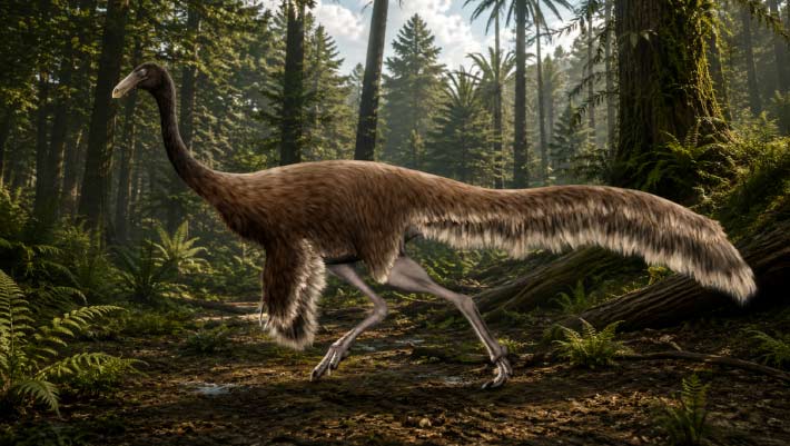
Rare Ostrich-Like Dinosaur Fossil Found on Canadian Island
Un rare fossile de dinosaure ressemblant à une autruche découvert sur une île canadienne
vertèbre Canada Colombie fossile Dinosauria Ornithomimosauria oiseau
Des paléontologues canadiens affirment avoir découvert une vertèbre caudale de dinosaure dans des roches marines vieilles de 75 à 80 millions d'années sur une petite île au large de la Colombie-Britannique, fournissant ainsi la preuve la plus claire à ce jour que des ornithomimosaures ressemblant à des oiseaux parcouraient autrefois l'ancienne côte du Pacifique de l'Amérique du Nord. L'article Un fossile rare de dinosaure ressemblant à une autruche trouvé sur une île canadienne est apparu en premier sur Sci.News : Breaking Science News.
25/05/2026 sci-news ⚙ Traduction automatique
Un ancien serpent de 50 pieds découvert en Inde pourrait être l'un des plus grands jamais vus
vertèbre Inde découverte
Un énorme serpent préhistorique découvert en Inde pourrait figurer parmi les plus grands serpents jamais rampés sur Terre. Nommé Vasuki indicus, cet ancien géant vivait il y a environ 47 millions d'années et on estime qu'il s'étendait sur une longueur étonnante de 11 à 15 mètres, rivalisant avec le légendaire Titanoboa. Des vertèbres fossilisées découvertes dans une mine de lignite du Gujarat révèlent un serpent au corps épais et puissant, probablement construit pour des embuscades lentes et furtives, semblable aux anacondas modernes.
29/04/2026 sciencedaily-paleo ⚙ Traduction automatique
1 2 3