Galerie d'images
Toutes les images de la base — taxons, formations et intervalles géologiques.
⚠ La fonctionnalité de récupération des images est en cours de test, des images non pertinentes peuvent apparaître.
2,093 image(s)

Photo of a skeletal mount of the incomplete holotype of the pachycephalosaur Homalocephale calathocercos in left lateral view, at the Mongolian Natural History Museum. Scan of physical photograph.


Obelignathus septimanicus holotype, MDE D30 from the 'Grès à Reptiles' Formation of Montouliers, southern France. Photographs and photogrammetric models of the right dentary in dorsal (A), anterior (B), medial (C), and lateral (D) views.

Right dentary in (A), medial; (B), dorsal; and (C) lateral views. Dashed black lines represent approximate contours of the missing areas. Dashed red lines indicate the distinctive banding pattern in the opal used to estimate the extent of the missing area. (D–F) Three-dimensional renders of the posterior dentary fragment in (D) lingual view showing erupted (blue) and developing germ teeth (pink); (E) Same as (D) but with dentary removed; (F) dorsal (occlusal) view of tooth row. (G-J) Three-dimensional render of the best-preserved tooth in (G) mesial, (H) lingual, (I) distal, and (J) labial views. (K) MicroCT scan of the posterior dentary fragment in axial view showing preservation of cancellous bone. Abbreviations: cab, cancellous bone; cr, tooth crown. Photo credit: Phil Bell.

Photo: mounted skeleton of Qantassaurus intrepidus at the Australian Museum, Sydney.


Skeletal elements of Lophorhothon atopus. (A, B and C) Partial skull roof and braincase (holotype FMNH 27383) in dorsal, ventral, and left lateral views. (D) Partial left nasal of FMNH 27383 in lateral view. (E) Left prefrontal of FMNH 27383 in lateral view. (F) Partial left jugal of FMNH 27383 in medial view. (G) Detail of marginal denticles of the dentary tooth in (H). (H) Apical half of a dentary tooth crown AUMP 2295 in lingual view. (I) Maxillary tooth crown of FMNH 27383 in labial view. (J) Left pubis of AUMP 2295 in lateral view. (K) Iliac process of the left ilium of FMNH 27383 in lateral view.

Illustration of the iberian iguanodontian Delapparentia turolensis.


The skull of the holotype of Acristavus, MOR 1155

Holotype skull of Augustynolophus morrisi (LACM 2852) on display at the Natural History Museum of Los Angeles County.

Huallasaurus (formerly Kritosaurus australis), Museo de Ciencias Naturales. CRISIS & REPRESENTACION (19 / 20 Diciembre 2011). crisisrepresentacion.wordpress.com/ Idea / Coordinación Loreto Garin Guzman + Federico Zukerfeld “Celebrando la caída de los Tiranosaurios” Performance con bandera en árabe-español: QUE SE VAYAN TODOS! En medio de dinosaurios y reliquias arqueológicas al interior de las galerías del Museo de Ciencias Naturales, celebramos la caída de los Tiranosaurio. A través de un picnic de conversación antropofágica, buscando interpretaciones y miradas comunes acerca de las transformaciones de los discursos y narrativas. Fotografía:Mathijs de Bruijne.


Reconstructed skull of Jaxartosaurus aralensis Riabinin, 1937. Only a posterior portion of the skull, as well as a surangular is known, with the rest of the skull being based mostly upon File:Aralosaurus skull.png. Jaxartosaurus is currently classified as a basal lambeosaurine, more derived than Aralosaurini, but more primitive than Parasaurolophini and Lambeosaurini. It might be more derived than Tsintaosaurini (Godefroit et al., 2004). References: Riabinin, A.M. (1937). "A New Finding of Dinosaurs in the Trans-Baikal Region". Ezhegodn. Vserossijskogo Palaeont. Obstcg. 11: 142–144 Godefroit, P.; Bolotsky, Y.L.; Van Itterbeeck, J. (2004). "The lambeosaurine dinosaur Amurosaurus riabinini, from the Maastrichtian of Far Eastern Russia". Acta Palaeontologica Polonica 49(4): 585–618

Barsboldia sicinskii gen. et sp.n., type specimen ZPAL MgD-11110, Nemegt Formation, N Nemegt, Nemegt Basin, Gobi Desert, Mongolia 1. Right lateral view of sacrum, neural spine of last dorsal and two anterior caudals included.


Yandusaurus hongheensis, a neornithischian dinosaur.


Partial skull of Manidens condorensis from the Middle Jurassic Cañadón Asfalto Formation of Argentina. Skull reconstructions in lateral view. Dashed lines indicate estimated edges. Abbreviations: a angular antfo antorbital fossa asaf anterior surangular foramen be buccal emargination bo basioccipital bt basal tubera d dentary d1, 2, 11 dentary tooth 1, 2, 11 emfo external mandibular fossa f frontal gl glenoid gr groove j jugal jfl jugal flange jh jugal horn m maxilla m1, 11 maxillary tooth 1, 11 n nasal pd predentary pm premaxilla po postorbital pof postorbital fossa popr paroccipital process q quadrate qj quadratojugal ri ridge sa surangular sq squamosal.

Kunbarrasaurus ieversi (QM F18101; holotype) skeleton in dorsal view. Scale = 20 cm.

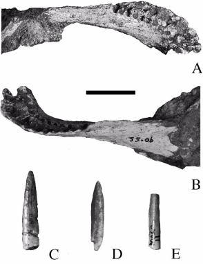
Figure 1, 1a. Outer and oral aspects of the imperfect dentary bone of Sarcolestes Leedsi, from the Oxford Clay of Peterborough. 2/3 nat size. s = symphysis. Figure 1b. A single tooth of the former. 3/1 nat size. Figure 2, 2a. Outer aspect and quadratic cavity of the hinder region of the same jaw. 2/3 nat size. Figure 3. A single tooth of Priodontognathus Phillipsi, 3/1 nat size, shown for purposes of comparison. Specimen in the Woodwardian Museum, Cambridge.

Cranial diagram of the ankylosaurine Tsagantegia longicranialis, holotype MPC 700/17 in A) dorsal and B) ventral views. Based on Tumanova 1993[1], Arbour & Currie 2015[2] and James I. Kirkland.[3] Abbreviations: asob: anterior supraorbital caputegulum - frca: frontal caputegulum - laca: lacrimal caputegulum - loca: loreal caputegulum - mso: middle supraorbital caputegulae - nasca: nasal caputegulum - nuca: nuchal caputegulum - poca: postocular caputegulum - prfca: prefrontal caputegulum - psob: posterior supraorbital caputegulum - qjh: quadratojugal horn - snca: supranarial caputegulum - sqh: squamosal horn

Skeletal mount of Gobisaurus domoculus on display at the Baoding Natural History Museum.


Hungarosaurus tormai (Ankylosauria, Nodosauridae), right dentary (MTM 2007.25.2) in lateral view


Vertebrae and spike of Lexovisaurus, Muséum national d'histoire naturelle, Paris.

Rendition of possible appearance of the dinosaur genus Nyasasaurus from the Middle Triassic, possibly the earliest known dinosaur. Black portions represent the partial skeletal fragments (a humerus and six vertebrae) from one specimen blue portions represent fragments from a second specimen (three cervical vertebrae) on which the current likely form of the animal is based.


Digital drawing of the known skeletal remains of Macrocollum itaquii. Known elements represented in white and unknown in gray.


Datousaurus. Fossil in Shanghai Science & Technology Museum

historická budova Národního muzea v Praze - západní dvorana s proskleným stropem a kostrou dinosaura / historic building of the National Museum in Prague - West hall with glass ceiling and dinosaur skeleton location: Prague, Czech Republic author: Jan Helebrant www.juhele.blogspot.com license CC0 Public Domain Dedication


Tastavinsaurus de El Castellar (Teruel, España). Fémur, tibia y fíbula izquierdos. En exposición en el museo paleontológico de Dinópolis.

Holotype vertebrae of Barrosasaurus casamiquelai, anterior dorsal MCF-PVPH-447/3 (left) and posterior dorsal MCF-PVPH-447/1 (right). Scale bar is 200 mm.

Figure 1: Geographic provenance and speculative reconstruction of the gigantic titanosaurian sauropod dinosaur Notocolossus gonzalezparejasi gen. et sp. nov. (a) Type locality of Notocolossus (indicated by star) in southern-most Mendoza Province, Argentina. (b) Reconstructed skeleton and body silhouette in right lateral view, with preserved elements of the holotype (UNCUYO-LD 301) in light green and those of the referred specimen (UNCUYO-LD 302) in orange. Scale bar, 1 m. (All images were hand drawn by the senior author [B.J.G.R.] and subsequently edited using Adobe Illustrator software.)

Skeleton of Gondwanatitan faustoi (MN 4111-V), not to scale.(adapted from Kellner and Campos, 2000).


Photographs and interpretive drawings of the titanosauroid Bonitasaura salgadoi Apesteguía, 2004 from the Upper Neuquén Group of Río Negro province, Patagonia, MPCA 460. A–D. Right dentary in dorsal (A), medial (B), ventral (C), and lateral (D) views. E. Isolated tooth in labial (E1) and lateral (E2) views; lingual view detail and schematic cross section showing hexagonal faceting (E3).


Paludititan nalatzensis (UBB NVM1-43), Maastrichtian, Haţeg Basin, Romania. Scale bar equals 10 cm.