Identifier: catalogueoffossi02bri (find matches)
Title: Catalogue of the fossil Reptilia and Amphibia in the British Museum (Natural history) ... By Richard Lydekker ..
Year: 1888 (1880s)
Authors: British Museum (Natural History). Dept. of Geology Lydekker, Richard, 1849-1915
Subjects: Reptiles, Fossil Amphibians, Fossil
Publisher: London, Printed by order of the Trustees
Contributing Library: Smithsonian Libraries
Digitizing Sponsor: Biodiversity Heritage Library
View Book Page: Book Viewer
About This Book: Catalog Entry
View All Images: All Images From Book
Click here to view book online to see this illustration in context in a browseable online version of this book.
Text Appearing Before Image:
pterygian; from theKimeridge Clay of Ely. \. a, proximal, b, distal epiphysis ; c, shaft. 46792. Two still smaller specimens; from the Kimeridge Clay ofDevizes, Wiltshire. Cunnington Collection. R. 400. Two phalangeals; from the Oxford Clay of Weymouth,Dorsetshire. Presented by C. Westendarp, Esq., 1884. 150 SAUEOPTERIGIA. R. 1381. One longitudinal half of the humerus or femur of a me- (Ficj.) dium-sized form, with the inner surface cut and polished; from the Kimeridge Clay of Ely. This specimen is figured in the woodcut on the preceding page, and shows the two epiphyses almost meeting in the middle of the shaft. No history. R. 1381 a. The proximal half of a larger humerus or femur, longitu-dinally bisected; from the Kimeridge Clay of Ely. Thecontour of the proximal epiphysis is well displayed. No history. 46912. The proximal portion of a still larger humerus or femur,longitudinally bisected; from Shotover. The whole ofthe proximal epiphysis is displayed, of which the terminal Kg. 47.
Text Appearing After Image:
Sauropterygian mandibles.—A. Peloneustesphilarchus; from the Oxford Clay.\. B. Thaitmatosaurus indicus; from the Upper Jurassic of India. ).0. Pksiosaimcs dolichodirus; from the Lower Lias. f. (From the Kec.Geol. Surv. Ind.) PLESI0SAURID2E. 151 extremity appears to have been separated by a small va-cuity from that of the distal epiphysis. No history. 42097. One lateral half of a humerus or femur, with the inner sur- face cut; from the Neocomian bone-bed of Potton, Bed-fordshire. The extremity of one epiphysis is entire anddetached from the shaft, while a section is shown of thatat the opposite end. Purchased, 1870. 42098. A small imperfect femur, with the proximal epiphysis de- tached and lying loose in the cup of the shaft; fromPotton. Purchased, 1870. Genus PELONEUSTES, Lydekker \ Skull and teeth of the general type of Pliosaurus, but the mandible(fig. 47, A) with a longer symphysis, which includes more than adozen teeth. Neck short, with the anterior vertebras relativelyshort. Ve
Note About Images
Please note that these images are extracted from scanned page images that may have been digitally enhanced for readability - coloration and appearance of these illustrations may not perfectly resemble the original work.


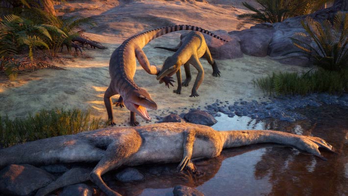
![Restoration of the spinosaurid dinosaur Siamosaurus in the Sao Khua Formation palaeoenvironment, with Sunosuchus in the middle left and a herd of Phuwiangosaurus in the background.
References:
Siamosaurus based on tooth specimens [1] and the neural spine of a possibly referable skeleton[2], with other missing elements filled in with relatives (Suchomimus[3], Baryonyx[4], IchthyovenatorFile:Ichthyovenator_laosensis_skeletal_reconstruction_by_PaleoGeek.png).
Phuwiangosaurus based on skeletal by Suteethorn et al. (2009)[5] and missing elements of skull of EuhelopusFile:Euhelopus.png.
Sunosuchus based on Suteethorn and Ingavat (1983)[6] and missing elements based on Goniopholis[7].](https://upload.wikimedia.org/wikipedia/commons/thumb/9/93/Sao_Khua_Formation_V2.png/330px-Sao_Khua_Formation_V2.png)


.jpg)