dent

Partie anatomique

59 image(s) · 34 Actualités

Galerie d'images

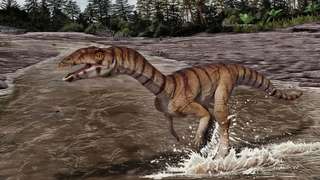
Restoration of the Chinese theropod "Sinopliosaurus fusuiensis" as a generalized spinosaurid. Silhouette of the holotype tooth is visible.
Taxons Sinopliosaurus

Restoration of the Chinese theropod "Sinopliosaurus fusuiensis" as a generalized spinosaurid. Silhouette of the holotype tooth is visible.

dent holotype Sinopliosaurus Spinosauridae
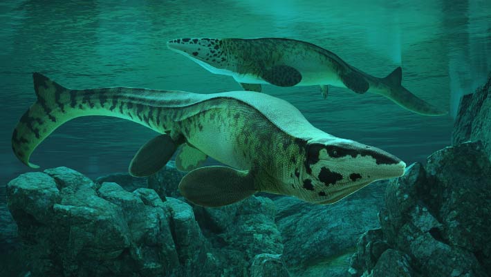
dent de Globidens aegyptiacus. La couronne dentaire montre bien le mode "broyeur" de ce Mosasaure.

dent de Globidens aegyptiacus. La couronne dentaire montre bien le mode "broyeur" de ce Mosasaure.

dent Globidens Igdamanosaurus Mosasaurus
dent de Globidens aegyptiacus. La couronne dentaire montre bien le mode "broyeur" chez ce Mosasaure. A noter la cavité, vide, d'une dent de remplacement.

dent de Globidens aegyptiacus. La couronne dentaire montre bien le mode "broyeur" chez ce Mosasaure. A noter la cavité, vide, d'une dent de remplacement.

dent Globidens Igdamanosaurus Mosasaurus
dent de Globidens aegyptiacus. La couronne dentaire montre bien le mode "broyeur" chez ce Mosasaure.

dent de Globidens aegyptiacus. La couronne dentaire montre bien le mode "broyeur" chez ce Mosasaure.

dent Globidens Igdamanosaurus Mosasaurus
dent de Globidens aegyptiacus et dent de Mosasaurus beaugei montrant les cavités de la dent de remplacement

dent de Globidens aegyptiacus et dent de Mosasaurus beaugei montrant les cavités de la dent de remplacement

dent Globidens Igdamanosaurus Mosasaurus
dent de Globidens aegyptiacus et dent de Mosasaurus beaugei montrant les cavités de la dent de remplacement
Taxons Igdamanosaurus

dent de Globidens aegyptiacus et dent de Mosasaurus beaugei montrant les cavités de la dent de remplacement

dent Globidens Igdamanosaurus Mosasaurus
Representative taxa from the early Campanian Muthmannsdorf fauna from the Grünbach Formation, eastern Austria. A Doratodon carcharidens (Mesoeucrocodylia) mandible (PIUW 2349/57) in dorsal view (photo by Márton Rabi) B Indeterminate azhdarchid (Pterosauria, Azhdarchidae), left humerus (PIUW 2349/102) in anterior view C ‘Megalosaurus pannoniensis’ basal tetanuran (Theropoda, Tetanurae), tooth (PIUW uncatalogued) in lateral view D Mochlodon suessi (Ornithopoda, Rhabdodontidae), right dentary (holotype, PIUW 2349/2) in medial view. Scale bars equal 2 cm in A, B and D and 1 cm in C.

Representative taxa from the early Campanian Muthmannsdorf fauna from the Grünbach Formation, eastern Austria. A Doratodon carcharidens (Mesoeucrocodylia) mandible (PIUW 2349/57) in dorsal view (photo by Márton Rabi) B Indeterminate azhdarchid (Pterosauria, Azhdarchidae), left humerus (PIUW 2349/102) in anterior view C ‘Megalosaurus pannoniensis’ basal tetanuran (Theropoda, Tetanurae), tooth (PIUW uncatalogued) in lateral view D Mochlodon suessi (Ornithopoda, Rhabdodontidae), right dentary (holotype, PIUW 2349/2) in medial view. Scale bars equal 2 cm in A, B and D and 1 cm in C.

humérus écaille dent Autriche +11
Representative taxa from the Santonian Iharkút fauna from the Csehbánya Formation, Bakony Mountains, western Hungary. A Pannoniasaurus inexpectatus (Squamata, Mosasauroidea), dorsal vertebra (MTM uncatalogued) in dorsal view (photo by Réka Kalmár) B Foxemys trabanti (Pleurodira, Bothremydidae), skull (MTM V 2010.215.1.) in dorsal view (photo by Márton Rabi). C Bicuspidon aff. hatzegiensis (Squamata, Borioteiioidea), left dentary (MTM 2006.112.1.) in medial view (photo by László Makádi) D Basal tetanuran (Theropoda, Tetanurae), tooth (MTM V.01.54) in ?lingual view E Indeterminate abelisaurid (Theropoda, Abelisauridae), pedal ungual phalanx (MTM V 2008.43.1.) in lateral view F Pneumatoraptor fodori (Theropoda, Paraves), left scapulocoracoid (holotype, MTM V 2008.38.1.) in lateral view G Mochlodon vorosi (Ornithopoda, Rhabdodontidae), left dentary (holotype, MTM V 2010.105.1) in lateral view H Bakonydraco galaczi (Pterosauria, Azhdarchidae), mandible (holotype, MTM 2007.110.1) in dorsal view I Iharkutosuchus makadii (Eusuchia, Hylaeochampsidae), skull (holotype, MTM 2006.52.1) in dorsal view J Hungarosaurus tormai (Ankylosauria, Nodosauridae), right dentary (MTM 2007.25.2) in lateral view K Bauxitornis mindszentyae (Aves, Enantiornithes), left tarsometatarsus (holotype, MTM V 2009.38.1) in anterior view L Ajkaceratops kozmai (Ceratopsia), fused rostral and premaxillae (holotype, MTM V 2009.192.1) in lateral view. Scale bars: 2 cm in A, V, G, H, I, J; 1 cm in D, E, F, K, L; 1 mm in C.

Representative taxa from the Santonian Iharkút fauna from the Csehbánya Formation, Bakony Mountains, western Hungary. A Pannoniasaurus inexpectatus (Squamata, Mosasauroidea), dorsal vertebra (MTM uncatalogued) in dorsal view (photo by Réka Kalmár) B Foxemys trabanti (Pleurodira, Bothremydidae), skull (MTM V 2010.215.1.) in dorsal view (photo by Márton Rabi). C Bicuspidon aff. hatzegiensis (Squamata, Borioteiioidea), left dentary (MTM 2006.112.1.) in medial view (photo by László Makádi) D Basal tetanuran (Theropoda, Tetanurae), tooth (MTM V.01.54) in ?lingual view E Indeterminate abelisaurid (Theropoda, Abelisauridae), pedal ungual phalanx (MTM V 2008.43.1.) in lateral view F Pneumatoraptor fodori (Theropoda, Paraves), left scapulocoracoid (holotype, MTM V 2008.38.1.) in lateral view G Mochlodon vorosi (Ornithopoda, Rhabdodontidae), left dentary (holotype, MTM V 2010.105.1) in lateral view H Bakonydraco galaczi (Pterosauria, Azhdarchidae), mandible (holotype, MTM 2007.110.1) in dorsal view I Iharkutosuchus makadii (Eusuchia, Hylaeochampsidae), skull (holotype, MTM 2006.52.1) in dorsal view J Hungarosaurus tormai (Ankylosauria, Nodosauridae), right dentary (MTM 2007.25.2) in lateral view K Bauxitornis mindszentyae (Aves, Enantiornithes), left tarsometatarsus (holotype, MTM V 2009.38.1) in anterior view L Ajkaceratops kozmai (Ceratopsia), fused rostral and premaxillae (holotype, MTM V 2009.192.1) in lateral view. Scale bars: 2 cm in A, V, G, H, I, J; 1 cm in D, E, F, K, L; 1 mm in C.

dent vertèbre Hongrie Santonien +14
Restoration of the spinosaurid dinosaur Siamosaurus in the Sao Khua Formation palaeoenvironment, with Sunosuchus in the middle left and a herd of Phuwiangosaurus in the background.
References:
Siamosaurus based on tooth specimens [1] and the neural spine of a possibly referable skeleton[2], with other missing elements filled in with relatives (Suchomimus[3], Baryonyx[4], IchthyovenatorFile:Ichthyovenator_laosensis_skeletal_reconstruction_by_PaleoGeek.png).
Phuwiangosaurus based on skeletal by Suteethorn et al. (2009)[5] and missing elements of skull of EuhelopusFile:Euhelopus.png.

Sunosuchus based on Suteethorn and Ingavat (1983)[6] and missing elements based on Goniopholis[7].
Formations Sao Khua

Restoration of the spinosaurid dinosaur Siamosaurus in the Sao Khua Formation palaeoenvironment, with Sunosuchus in the middle left and a herd of Phuwiangosaurus in the background. References: Siamosaurus based on tooth specimens [1] and the neural spine of a possibly referable skeleton[2], with other missing elements filled in with relatives (Suchomimus[3], Baryonyx[4], IchthyovenatorFile:Ichthyovenator_laosensis_skeletal_reconstruction_by_PaleoGeek.png). Phuwiangosaurus based on skeletal by Suteethorn et al. (2009)[5] and missing elements of skull of EuhelopusFile:Euhelopus.png. Sunosuchus based on Suteethorn and Ingavat (1983)[6] and missing elements based on Goniopholis[7].

dent dessin Sao Khua spécimen +11
A 1.7cm tall tooth from Liodon anceps.  Cretaceous, Phosphate beds, Kouribga, Morocco.

A 1.7cm tall tooth from Liodon anceps. Cretaceous, Phosphate beds, Kouribga, Morocco.

dent Maroc Crétacé Leiodon +1
Fig. 2. Side view of crown of tooth of Cardiodon rugulosus.
Fig. 3. Fore end of the same tooth.
Fig. 4. Hind end of the crown of another tooth of Cardiodon rugulosus.
Fig. 5. Magnified view of markings on the surface of the enamel of the same tooth.
All the figures are of the natural size.

2-5 are from the Forest Marble of Bradford, Wilts. In the Collection of Channing Pearce, Esq., of that town.
Taxons Cardiodon

Fig. 2. Side view of crown of tooth of Cardiodon rugulosus. Fig. 3. Fore end of the same tooth. Fig. 4. Hind end of the crown of another tooth of Cardiodon rugulosus. Fig. 5. Magnified view of markings on the surface of the enamel of the same tooth. All the figures are of the natural size. 2-5 are from the Forest Marble of Bradford, Wilts. In the Collection of Channing Pearce, Esq., of that town.

dent Cardiodon
Fig. 1. Type material of Amygdalodon patagonicus Cabrera. 1947. A. B. Tooth, MLP 46-VIII-21-1/13, in lingual (A) and labial (B) view. C. D, E. Cervical vertebra, MLP 46-VIII-21-1/8. in right lateral (C, stereopair), ventral (D) and left lateral (E) view. F, G. Right cervical prezygapophysis, MLP 46-VIII-21-117. in dorsal (F) and medial (G) view. H. Anterior dorsal neural spine, MLP 46-VIII-21-116. in posterior view. I, J, K. Posterior dorsal vertebra, MLP 46-VIII-21-112, lectotype, in right lateral view (I, stereopair), right lateral view, with right wall of neural canal remived (J), and posterior (K) view. L, M. Caudal vertebra, MLP 46-VIII-21-1/3, in left lateral (L) and posterior (M) view. N. Caudal vertebra, MLP 46-VIII-21-1/4, in right lateral view. (O). Proximal fragment of a dorsal rib, MLP 46-VIII-21-1/9, in anterolateral view. Abbreviations: acdl, anterior centrodiapophyseal lamina; cprl, centroprezygapophyseal lamina; g, groove; k, ventral keel; pcdl, posterior centrodiapophyseal lamina; pod, postzygodiapophyseal lamina; poz, postzygapophysis; pp, parapophysis; prdl, prezygodiapophyseal lamina; spol, spinopostzgapophyseal lamina; sprl, spinoprezygapophyseal lamina; tprl, intraprezygapophyseal lamina. Scale bars 1 cm (A, B) and 10 cm (C-0).
Taxons Amygdalodon

Fig. 1. Type material of Amygdalodon patagonicus Cabrera. 1947. A. B. Tooth, MLP 46-VIII-21-1/13, in lingual (A) and labial (B) view. C. D, E. Cervical vertebra, MLP 46-VIII-21-1/8. in right lateral (C, stereopair), ventral (D) and left lateral (E) view. F, G. Right cervical prezygapophysis, MLP 46-VIII-21-117. in dorsal (F) and medial (G) view. H. Anterior dorsal neural spine, MLP 46-VIII-21-116. in posterior view. I, J, K. Posterior dorsal vertebra, MLP 46-VIII-21-112, lectotype, in right lateral view (I, stereopair), right lateral view, with right wall of neural canal remived (J), and posterior (K) view. L, M. Caudal vertebra, MLP 46-VIII-21-1/3, in left lateral (L) and posterior (M) view. N. Caudal vertebra, MLP 46-VIII-21-1/4, in right lateral view. (O). Proximal fragment of a dorsal rib, MLP 46-VIII-21-1/9, in anterolateral view. Abbreviations: acdl, anterior centrodiapophyseal lamina; cprl, centroprezygapophyseal lamina; g, groove; k, ventral keel; pcdl, posterior centrodiapophyseal lamina; pod, postzygodiapophyseal lamina; poz, postzygapophysis; pp, parapophysis; prdl, prezygodiapophyseal lamina; spol, spinopostzgapophyseal lamina; sprl, spinoprezygapophyseal lamina; tprl, intraprezygapophyseal lamina. Scale bars 1 cm (A, B) and 10 cm (C-0).

dent vertèbre Amygdalodon
Right dentary in (A), medial; (B), dorsal; and (C) lateral views. Dashed black lines represent approximate contours of the missing areas. Dashed red lines indicate the distinctive banding pattern in the opal used to estimate the extent of the missing area. (D–F) Three-dimensional renders of the posterior dentary fragment in (D) lingual view showing erupted (blue) and developing germ teeth (pink); (E) Same as (D) but with dentary removed; (F) dorsal (occlusal) view of tooth row. (G-J) Three-dimensional render of the best-preserved tooth in (G) mesial, (H) lingual, (I) distal, and (J) labial views. (K) MicroCT scan of the posterior dentary fragment in axial view showing preservation of cancellous bone. Abbreviations: cab, cancellous bone; cr, tooth crown. Photo credit: Phil Bell.
Taxons Weewarrasaurus

Right dentary in (A), medial; (B), dorsal; and (C) lateral views. Dashed black lines represent approximate contours of the missing areas. Dashed red lines indicate the distinctive banding pattern in the opal used to estimate the extent of the missing area. (D–F) Three-dimensional renders of the posterior dentary fragment in (D) lingual view showing erupted (blue) and developing germ teeth (pink); (E) Same as (D) but with dentary removed; (F) dorsal (occlusal) view of tooth row. (G-J) Three-dimensional render of the best-preserved tooth in (G) mesial, (H) lingual, (I) distal, and (J) labial views. (K) MicroCT scan of the posterior dentary fragment in axial view showing preservation of cancellous bone. Abbreviations: cab, cancellous bone; cr, tooth crown. Photo credit: Phil Bell.

os dent Weewarrasaurus
Skeletal elements of Lophorhothon atopus. (A, B and C) Partial skull roof and braincase (holotype FMNH 27383) in dorsal, ventral, and left lateral views. (D) Partial left nasal of FMNH 27383 in lateral view. (E) Left prefrontal of FMNH 27383 in lateral view. (F) Partial left jugal of FMNH 27383 in medial view. (G) Detail of marginal denticles of the dentary tooth in (H). (H) Apical half of a dentary tooth crown AUMP 2295 in lingual view. (I) Maxillary tooth crown of FMNH 27383 in labial view. (J) Left pubis of AUMP 2295 in lateral view. (K) Iliac process of the left ilium of FMNH 27383 in lateral view.
Taxons Lophorhothon

Skeletal elements of Lophorhothon atopus. (A, B and C) Partial skull roof and braincase (holotype FMNH 27383) in dorsal, ventral, and left lateral views. (D) Partial left nasal of FMNH 27383 in lateral view. (E) Left prefrontal of FMNH 27383 in lateral view. (F) Partial left jugal of FMNH 27383 in medial view. (G) Detail of marginal denticles of the dentary tooth in (H). (H) Apical half of a dentary tooth crown AUMP 2295 in lingual view. (I) Maxillary tooth crown of FMNH 27383 in labial view. (J) Left pubis of AUMP 2295 in lateral view. (K) Iliac process of the left ilium of FMNH 27383 in lateral view.

dent holotype Lophorhothon partiel +1
Partial skull of Manidens condorensis from the Middle Jurassic Cañadón Asfalto Formation of Argentina. Skull reconstructions in lateral view. Dashed lines indicate estimated edges. Abbreviations: a angular antfo antorbital fossa asaf anterior surangular foramen be buccal emargination bo basioccipital bt basal tubera d dentary d1, 2, 11 dentary tooth 1, 2, 11 emfo external mandibular fossa f frontal gl glenoid gr groove j jugal jfl jugal flange jh jugal horn m maxilla m1, 11 maxillary tooth 1, 11 n nasal pd predentary pm premaxilla po postorbital pof postorbital fossa popr paroccipital process q quadrate qj quadratojugal ri ridge sa surangular sq squamosal.

Partial skull of Manidens condorensis from the Middle Jurassic Cañadón Asfalto Formation of Argentina. Skull reconstructions in lateral view. Dashed lines indicate estimated edges. Abbreviations: a angular antfo antorbital fossa asaf anterior surangular foramen be buccal emargination bo basioccipital bt basal tubera d dentary d1, 2, 11 dentary tooth 1, 2, 11 emfo external mandibular fossa f frontal gl glenoid gr groove j jugal jfl jugal flange jh jugal horn m maxilla m1, 11 maxillary tooth 1, 11 n nasal pd predentary pm premaxilla po postorbital pof postorbital fossa popr paroccipital process q quadrate qj quadratojugal ri ridge sa surangular sq squamosal.

dent Argentine Jurassique Jurassique moyen +4
Figure 1, 1a. Outer and oral aspects of the imperfect dentary bone of Sarcolestes Leedsi, from the Oxford Clay of Peterborough. 2/3 nat size. s = symphysis. Figure 1b. A single tooth of the former. 3/1 nat size. Figure 2, 2a. Outer aspect and quadratic cavity of the hinder region of the same jaw. 2/3 nat size. Figure 3. A single tooth of Priodontognathus Phillipsi, 3/1 nat size, shown for purposes of comparison. Specimen in the Woodwardian Museum, Cambridge.

Figure 1, 1a. Outer and oral aspects of the imperfect dentary bone of Sarcolestes Leedsi, from the Oxford Clay of Peterborough. 2/3 nat size. s = symphysis. Figure 1b. A single tooth of the former. 3/1 nat size. Figure 2, 2a. Outer aspect and quadratic cavity of the hinder region of the same jaw. 2/3 nat size. Figure 3. A single tooth of Priodontognathus Phillipsi, 3/1 nat size, shown for purposes of comparison. Specimen in the Woodwardian Museum, Cambridge.

os dent musée Oxford Clay +3
1 2 3 4

Actualités

Une « plante grimpante des sables » géante vieille de 240 millions d’années découverte cachée dans un mur de soutènement
dent prédateur Australie fossile découverte squelette
Un fossile oublié caché dans un mur de jardin s’est avéré être l’une des découvertes préhistoriques les plus remarquables d’Australie. Les scientifiques ont maintenant identifié l'amphibien Arenaerpeton supinatus, vieux de 240 millions d'années, révélant un squelette presque parfaitement conservé, avec de rares traces de peau. Cet ancien prédateur de rivière, mesurant environ 1,2 mètre de long, ressemblait un peu à une salamandre géante, mais il était plus volumineux et armé de redoutables dents en forme de crocs.
06/05/2026 sciencedaily ⚙ Traduction automatique
Early Platypuses Had Strong Teeth and Powerful Jaws, Fossils Show
Les premiers ornithorynques avaient des dents solides et des mâchoires puissantes, selon des fossiles
mâchoire dent Australie fossile formation
De nouveaux fossiles de la Formation de Namba, en Australie méridionale, révèlent qu'il y a 25 millions d'années, Obdurodon insignis prospérait dans les lacs intérieurs luxuriants aux côtés de dauphins d'eau douce et d'autres espèces aujourd'hui disparues. L'article Les premiers ornithorynques avaient des dents solides et des mâchoires puissantes, Fossils Show est apparu en premier sur Sci.News : Breaking Science News.
28/04/2026 sci-news ⚙ Traduction automatique
La « plus vieille pieuvre » du monde n’a jamais été une pieuvre
dent fossile découverte évolution
Un célèbre fossile du « plus vieux poulpe » a été exposé comme un cas d’identité erronée. Une imagerie avancée a révélé des dents cachées, montrant qu'il s'agissait en réalité d'un nautile et non d'une pieuvre. La confusion venait de la décomposition qui avait modifié sa forme avant la fossilisation. Cette découverte réécrit une partie de l’histoire de l’évolution, repoussant la véritable origine des poulpes bien plus tard dans le temps.
07/04/2026 sciencedaily ⚙ Traduction automatique
Des dents vieilles de 2 700 ans révèlent la vie cachée des Italiens de l'âge du fer
dent alimentation croissance Italie
Les dents de l’âge du fer du sud de l’Italie sont devenues des capsules temporelles, préservant les détails intimes de l’enfance et de l’alimentation. Les lignes de croissance dans l'émail révèlent des moments de stress au début de la vie, tandis que la plaque durcie contient des restes microscopiques de céréales, de légumineuses et d'aliments fermentés. Les résultats suggèrent une communauté dotée de ressources alimentaires diverses et de solides liens méditerranéens. Même un petit échantillon offre un aperçu saisissant de la façon dont les gens vivaient, grandissaient et mangeaient il y a près de trois millénaires.
04/03/2026 sciencedaily ⚙ Traduction automatique
Cet animal ancien fut l'un des premiers à manger des plantes sur terre
dent fossile crâne
Il y a des centaines de millions d’années, les premiers animaux à ramper sur terre étaient de purs mangeurs de viande, alors même que les plantes avaient déjà envahi le paysage. Aujourd’hui, les scientifiques ont découvert un fossile vieux de 307 millions d’années qui réécrit cette histoire : l’un des premiers vertébrés terrestres connus à avoir commencé à manger des plantes. L'animal, nommé Tyrannoroter heberti, était une créature trapue de la taille d'un ballon de football avec un crâne rempli de dents spécialisées conçues pour écraser et broyer la végétation.
11/02/2026 sciencedaily ⚙ Traduction automatique
1 2 3 4 5 6 7