←

squelette

Type de vue

162 image(s) · 26 Actualités

Galerie d'images

Skeleton of Tehuelchesaurus at the Museo Paleontológico Egidio Feruglio in Trelew, Argentina
Taxons Tehuelchesaurus

Skeleton of Tehuelchesaurus at the Museo Paleontológico Egidio Feruglio in Trelew, Argentina

Argentine Tehuelchesaurus squelette
FSAC-OB 232, holotype skeleton of Barbaridactylus grandis.
(A) Left humerus in anterior view, (B) right femur in anterior view, (C) right radius and ulna in posterior view, (D) cervical vertebra in ventral view, (E) left scapulocoracoid in medial view, and (F) posterior ramus of the right mandible in medial view.

Abbreviations: co, coracoid; cot, cotyle of mandible; cvr, cervical ribs; dm, dorsal margin of mandible; dpc, deltopectoral crest; gl, glenoid; fh, femoral head; gt, greater trochanter; hh, humeral head; hyp, hypapophysis; olp, olecranon process; pex, postexapophysis; pn, pneumatopore; rad, radius; sca, scapula; scp, supracondylar process of the ectepicondyle; uc, ulnar crest; uln, ulna; vm, ventral margin of mandible.
Taxons Barbaridactylus

FSAC-OB 232, holotype skeleton of Barbaridactylus grandis. (A) Left humerus in anterior view, (B) right femur in anterior view, (C) right radius and ulna in posterior view, (D) cervical vertebra in ventral view, (E) left scapulocoracoid in medial view, and (F) posterior ramus of the right mandible in medial view. Abbreviations: co, coracoid; cot, cotyle of mandible; cvr, cervical ribs; dm, dorsal margin of mandible; dpc, deltopectoral crest; gl, glenoid; fh, femoral head; gt, greater trochanter; hh, humeral head; hyp, hypapophysis; olp, olecranon process; pex, postexapophysis; pn, pneumatopore; rad, radius; sca, scapula; scp, supracondylar process of the ectepicondyle; uc, ulnar crest; uln, ulna; vm, ventral margin of mandible.

crête humérus vertèbre holotype +2
Nemicolopterus crypticus holotype (IVPP V-14377).

(A) Skeleton overview, and (B) schematic drawing. (C) Skull (right lateral view), and (D) schematic drawing. Abbreviations: cdv, caudal vertebrae; co, coracoid; cv, cervical vertebra; d1–d4, digits 1–4; f, frontal; fe, femur; h, humerus; hy, hyoid; il, illium; is, ischium; j, jugal; l, left; la, lacrimal; mand, mandible; mc, metacarpal; mt, metatarsal; naof, nasoantorbital fenestra; or, orbit; pa, parietal; pm, premaxilla; ph, phalanx; ti, tibia; r, right; sca, scapula. Scale bars: A–B, 100 mm; C–D, 5 mm.
Taxons Nemicolopterus

Nemicolopterus crypticus holotype (IVPP V-14377). (A) Skeleton overview, and (B) schematic drawing. (C) Skull (right lateral view), and (D) schematic drawing. Abbreviations: cdv, caudal vertebrae; co, coracoid; cv, cervical vertebra; d1–d4, digits 1–4; f, frontal; fe, femur; h, humerus; hy, hyoid; il, illium; is, ischium; j, jugal; l, left; la, lacrimal; mand, mandible; mc, metacarpal; mt, metatarsal; naof, nasoantorbital fenestra; or, orbit; pa, parietal; pm, premaxilla; ph, phalanx; ti, tibia; r, right; sca, scapula. Scale bars: A–B, 100 mm; C–D, 5 mm.

humérus écaille vertèbre dessin +4
Skeleton of Bagualia at the Museo Paleontológico Egidio Feruglio in Trelew, Argentina
Taxons Bagualia

Skeleton of Bagualia at the Museo Paleontológico Egidio Feruglio in Trelew, Argentina

Argentine Bagualia squelette
Reconstructed skeleton of Pterorhynchus wellnhoferi
Taxons Pterorhynchus

Reconstructed skeleton of Pterorhynchus wellnhoferi

Pterorhynchus squelette
Squelette partiel de Wulatesaurus.
Taxons Bannykus

Squelette partiel de Wulatesaurus.

Japon Bannykus partiel squelette
Xianshanosaurus skeleton
Taxons Xianshanosaurus

Xianshanosaurus skeleton

Xianshanosaurus squelette
Skeleton mount of Dallasaurus turneri at the Perot Museum
Taxons Dallasaurus

Skeleton mount of Dallasaurus turneri at the Perot Museum

musée Dallasaurus squelette
Reconstruction of the terrestrial paleoenvironmental setting of the Sao Khua Formation by Renata Cunha.
In the center, a generalized spinosaurid feeds on a sauropod. This trophic relationship is hypothesized based on isolated tooth crowns found in association with a sauropod skeleton [67]. In the background, a small pack of the ornithomimosaur theropod Kinnareemimus. Both sauropods and ornithomimosaurs (as part of the “herbivorous” theropods) were found to be positively associated with terrestrial paleoenvironments by Butler and Barrett [15].

(cropped from File:Spinosaurid and Kinnareemimus.PNG)
Taxons Kinnareemimus

Reconstruction of the terrestrial paleoenvironmental setting of the Sao Khua Formation by Renata Cunha. In the center, a generalized spinosaurid feeds on a sauropod. This trophic relationship is hypothesized based on isolated tooth crowns found in association with a sauropod skeleton [67]. In the background, a small pack of the ornithomimosaur theropod Kinnareemimus. Both sauropods and ornithomimosaurs (as part of the “herbivorous” theropods) were found to be positively associated with terrestrial paleoenvironments by Butler and Barrett [15]. (cropped from File:Spinosaurid and Kinnareemimus.PNG)

dent Sao Khua Kinnareemimus Ornithomimosauria +3
Alvarezsaurus skeleton restoration, Copenhagen.
Taxons Alvarezsaurus

Alvarezsaurus skeleton restoration, Copenhagen.

Alvarezsauria squelette
Skeletons of Bonatitan and Austroraptor at the Museo Argentino de Ciencias Naturales Bernardino Rivadavia
Taxons Bonatitan

Skeletons of Bonatitan and Austroraptor at the Museo Argentino de Ciencias Naturales Bernardino Rivadavia

Austroraptor Bonatitan squelette
Reconstructed skeleton of Daxiatitan binglingi.
Taxons Daxiatitan

Reconstructed skeleton of Daxiatitan binglingi.

Daxiatitan squelette
Identifier: waterreptilesofp1914will
Title: Water reptiles of the past and present
Year: 1914 (1910s)
Authors:  Williston, Samuel Wendell, 1851-1918
Subjects:  Aquatic reptiles
Publisher:  Chicago, Ill., The University of Chicago Press
Contributing Library:  Boston Public Library
Digitizing Sponsor:  Boston Public Library

View Book Page: Book Viewer
About This Book: Catalog Entry
View All Images: All Images From Book
Click here to view book online to see this illustration in context in a browseable online version of this book.

Text Appearing Before Image:
served and very complete skeletons ofdifferent species of ichthyosaurs from the Jurassic deposits ofWiirtemberg, in which remains of these animals occur in great 112 WATER REPTILES OF THE PAST AND PRESENT profusion. His researches, and those of several authors since then,supplementing and confirming or disproving those of the manyobservers made during the preceding seventy years, have finallydetermined almost perfectly the complete structure of the moretypical ichthyosaurs, enabling us to infer not a little as to theirhabits and distribution in the old Jurassic oceans. Within thepast few years the discoveries of Professor J. C. Merriam of Cali-fornia have likewise added greatly to our knowledge of the earlierichthyosaurs. It may now truthfully be said that of no group ofextinct reptiles do we have a more complete and satisfactory knowl-edge than of the ichthyosaurs. Nevertheless we have yet very much more to learn about theorder Ichthyosauria as a whole—whence they came and how they
Text Appearing After Image:
Fig. 52.—Ichthyosaurus quadricissus.museum, from Dr. Dreverman. Photograph of specimen in Senckenberg originated; what their nearest kin were among other reptiles; andespecially, more about the connecting links between them andterrestrial reptiles. They have, as an order, so isolated a position,are so widely separated from all other reptiles in structure, that theyhave long been a puzzle to paleontologists. Like the whales andother cetaceans among mammals, we know the ichthyosaurs wellin the plenitude of their power and the fulness of their development,but have yet only an imperfect knowledge of their earlier history,and none whatever of their earliest. However, as will be seenfarther on, the recent discoveries by Merriam have shed much lighton some of the stages of their evolution. So nearly perfectly wereall the later ichthyosaurs adapted to their life in the water that itwas believed by nearly all paleontologists until about a score of years ICHTHYOSAURIA 3 ago that they had desc

Note About Images

Please note that these images are extracted from scanned page images that may have been digitally enhanced for readability - coloration and appearance of these illustrations may not perfectly resemble the original work.
Taxons Stenopterygiidae

Identifier: waterreptilesofp1914will Title: Water reptiles of the past and present Year: 1914 (1910s) Authors: Williston, Samuel Wendell, 1851-1918 Subjects: Aquatic reptiles Publisher: Chicago, Ill., The University of Chicago Press Contributing Library: Boston Public Library Digitizing Sponsor: Boston Public Library View Book Page: Book Viewer About This Book: Catalog Entry View All Images: All Images From Book Click here to view book online to see this illustration in context in a browseable online version of this book. Text Appearing Before Image: served and very complete skeletons ofdifferent species of ichthyosaurs from the Jurassic deposits ofWiirtemberg, in which remains of these animals occur in great 112 WATER REPTILES OF THE PAST AND PRESENT profusion. His researches, and those of several authors since then,supplementing and confirming or disproving those of the manyobservers made during the preceding seventy years, have finallydetermined almost perfectly the complete structure of the moretypical ichthyosaurs, enabling us to infer not a little as to theirhabits and distribution in the old Jurassic oceans. Within thepast few years the discoveries of Professor J. C. Merriam of Cali-fornia have likewise added greatly to our knowledge of the earlierichthyosaurs. It may now truthfully be said that of no group ofextinct reptiles do we have a more complete and satisfactory knowl-edge than of the ichthyosaurs. Nevertheless we have yet very much more to learn about theorder Ichthyosauria as a whole—whence they came and how they Text Appearing After Image: Fig. 52.—Ichthyosaurus quadricissus.museum, from Dr. Dreverman. Photograph of specimen in Senckenberg originated; what their nearest kin were among other reptiles; andespecially, more about the connecting links between them andterrestrial reptiles. They have, as an order, so isolated a position,are so widely separated from all other reptiles in structure, that theyhave long been a puzzle to paleontologists. Like the whales andother cetaceans among mammals, we know the ichthyosaurs wellin the plenitude of their power and the fulness of their development,but have yet only an imperfect knowledge of their earlier history,and none whatever of their earliest. However, as will be seenfarther on, the recent discoveries by Merriam have shed much lighton some of the stages of their evolution. So nearly perfectly wereall the later ichthyosaurs adapted to their life in the water that itwas believed by nearly all paleontologists until about a score of years ICHTHYOSAURIA 3 ago that they had desc Note About Images Please note that these images are extracted from scanned page images that may have been digitally enhanced for readability - coloration and appearance of these illustrations may not perfectly resemble the original work.

livre musée Jurassique spécimen +7
Identifier: annalsofmedicalh01pack (find matches)
Title: Annals of medical history
Year: 1917 (1910s)
Authors:  Packard, Francis R. (Francis Randolph), 1870-1950
Subjects:  Medicine
Publisher:  New York P.B. Hoeber
Contributing Library:  Gerstein - University of Toronto
Digitizing Sponsor:  University of Toronto

View Book Page: Book Viewer
About This Book: Catalog Entry
View All Images: All Images From Book
Click here to view book online to see this illustration in context in a browseable online version of this book.

Text Appearing Before Image:
ail strongly Hexed, and the toescontracted and appressed. The whole atti-tude strongly suggests a spastic distress,possibly brought on by some form ofpoisoning of the central nervous system,from infection or the deglutition of somepoisonous substance. 14. Osteomalacia is evidently the causeof the hypertrophy of the bones of Lim-nocyron potens, an early carnivore from theWashakie Eocene of Wyoming, nearly3,000,000 years old. MATERIALS AND METHODS The material described in the presentpaper has been loaned the writer lor de-scription by the Field Museum of Chii Osborn: Hull. Amcr. \lus. Natl. Hist., 1917,vol. 35. !>• 733. P>- 28. Studies in Paleopathology 393 by the American Museum of Natural His-tory of New York City, by Walker Museumof the University of Chicago, and by the Uni-versity of Kansas Natural History Museum. made by the well-known petrographic meth-ods so common in all geological labora-tories. The diagnoses, where they are at-tempted, arc made from comparisons of the
Text Appearing After Image:
Fig. 20. The skeleton of Strutbiomimus altus, a small dinosaur from the BeliyRiver series (Cretaceous), Red Deer River, Alberta, Canada, now regarded as ofapproximately the same age as the Judith River series. The unique feature ofthe skull is the total absence of teeth, with a size of skull one-third larger thanthe ostrich and a length of body of about fifteen feet. The position of the skeletonis decidedly that of the opisthotonos which may be regarded as an indication ofdisease. (After Osborn). A beautiful specimen of an osteoma, theonly one known so far, on the vertebra of aKansas Cretaceous mosasaur, was given thewriter by Dr. J. M. Armstrong of St. Paul.The writer expresses his obligations to thegentlemen connected with the above-men-tioned institutions and to Dr. Armstrong.The methods used are a combination ofprocedures in the various lines involved.Microscopic sections, which can be madethin enough for immersion lens study, are material with similar lesions in recent hu-man mat

Note About Images

Please note that these images are extracted from scanned page images that may have been digitally enhanced for readability - coloration and appearance of these illustrations may not perfectly resemble the original work.
Taxons Hexing

Identifier: annalsofmedicalh01pack (find matches) Title: Annals of medical history Year: 1917 (1910s) Authors: Packard, Francis R. (Francis Randolph), 1870-1950 Subjects: Medicine Publisher: New York P.B. Hoeber Contributing Library: Gerstein - University of Toronto Digitizing Sponsor: University of Toronto View Book Page: Book Viewer About This Book: Catalog Entry View All Images: All Images From Book Click here to view book online to see this illustration in context in a browseable online version of this book. Text Appearing Before Image: ail strongly Hexed, and the toescontracted and appressed. The whole atti-tude strongly suggests a spastic distress,possibly brought on by some form ofpoisoning of the central nervous system,from infection or the deglutition of somepoisonous substance. 14. Osteomalacia is evidently the causeof the hypertrophy of the bones of Lim-nocyron potens, an early carnivore from theWashakie Eocene of Wyoming, nearly3,000,000 years old. MATERIALS AND METHODS The material described in the presentpaper has been loaned the writer lor de-scription by the Field Museum of Chii Osborn: Hull. Amcr. \lus. Natl. Hist., 1917,vol. 35. !>• 733. P>- 28. Studies in Paleopathology 393 by the American Museum of Natural His-tory of New York City, by Walker Museumof the University of Chicago, and by the Uni-versity of Kansas Natural History Museum. made by the well-known petrographic meth-ods so common in all geological labora-tories. The diagnoses, where they are at-tempted, arc made from comparisons of the Text Appearing After Image: Fig. 20. The skeleton of Strutbiomimus altus, a small dinosaur from the BeliyRiver series (Cretaceous), Red Deer River, Alberta, Canada, now regarded as ofapproximately the same age as the Judith River series. The unique feature ofthe skull is the total absence of teeth, with a size of skull one-third larger thanthe ostrich and a length of body of about fifteen feet. The position of the skeletonis decidedly that of the opisthotonos which may be regarded as an indication ofdisease. (After Osborn). A beautiful specimen of an osteoma, theonly one known so far, on the vertebra of aKansas Cretaceous mosasaur, was given thewriter by Dr. J. M. Armstrong of St. Paul.The writer expresses his obligations to thegentlemen connected with the above-men-tioned institutions and to Dr. Armstrong.The methods used are a combination ofprocedures in the various lines involved.Microscopic sections, which can be madethin enough for immersion lens study, are material with similar lesions in recent hu-man mat Note About Images Please note that these images are extracted from scanned page images that may have been digitally enhanced for readability - coloration and appearance of these illustrations may not perfectly resemble the original work.

os vertèbre livre musée +11
Photo of the partial holotype skeleton of the ornithomimosaur Garudimimus brevipes, as mounted in the Mongolian Natural History Museum. Scan of physical photograph.
Taxons Garudimimus

Photo of the partial holotype skeleton of the ornithomimosaur Garudimimus brevipes, as mounted in the Mongolian Natural History Museum. Scan of physical photograph.

musée holotype Garudimimus Ornithomimosauria +2
Selected elements used in the diagnosis of Saltriovenator zanellai n. gen. n. sp.

Right humerus in medial (A), frontal (B) and distal (C) views; (D) left scapula, medial view; (E) right scapular glenoid and coracoid, lateral view; (F) furcula, ventral view; tooth, labial (G) and apical (H) views; (I) left humerus, medial view; right second metacarpal in dorsal (J), lateral (L) and distal (N) views; first phalanx of the right second digit in dorsal (K), lateral (M) and proximal (O) views; (P–T) right third digit in proximal, dorsal and lateral views; (U) right distal tarsal IV, proximal view; third right metatarsal in proximal (V) and frontal (X) views; second right metatarsal, proximal (W) and frontal (Y) views; (Z) reconstructed skeleton showing identified elements (red). Abbreviations as in text, asterisks mark autapomorphic traits. Scale bars: 10 cm in (A)–(E), (I), and (U)–(Y); two cm in (F), and (J)–(T); one cm in (G).
Taxons Saltriovenator

Selected elements used in the diagnosis of Saltriovenator zanellai n. gen. n. sp. Right humerus in medial (A), frontal (B) and distal (C) views; (D) left scapula, medial view; (E) right scapular glenoid and coracoid, lateral view; (F) furcula, ventral view; tooth, labial (G) and apical (H) views; (I) left humerus, medial view; right second metacarpal in dorsal (J), lateral (L) and distal (N) views; first phalanx of the right second digit in dorsal (K), lateral (M) and proximal (O) views; (P–T) right third digit in proximal, dorsal and lateral views; (U) right distal tarsal IV, proximal view; third right metatarsal in proximal (V) and frontal (X) views; second right metatarsal, proximal (W) and frontal (Y) views; (Z) reconstructed skeleton showing identified elements (red). Abbreviations as in text, asterisks mark autapomorphic traits. Scale bars: 10 cm in (A)–(E), (I), and (U)–(Y); two cm in (F), and (J)–(T); one cm in (G).

humérus écaille dent dessin +2
1 2 3 4 5 6 7 8 9 10 11

Actualités

Un fossile remarquable révèle le régime alimentaire d'un ichtyosaure du Jurassique supérieur
Allemagne Jurassique Jurassique supérieur fossile spécimen Aegirosaurus Ichthyosauria Jabalisaurus squelette
Les scientifiques étudiant l’ichtyosaure Jabalisaurus tethyensis, récemment décrit, ont acquis un aperçu remarquable de son régime alimentaire. Un fossile magnifiquement conservé du sud de l'Allemagne contient des preuves des derniers repas de l'animal. De plus, le spécimen préserve des détails importants du squelette et des contours du corps. Le fossile, numéro de spécimen JME-SOS-08369, était auparavant attribué à Aegirosaurus. Cependant, les chercheurs
24/08/2026 everythingdinosaur ⚙ Traduction automatique
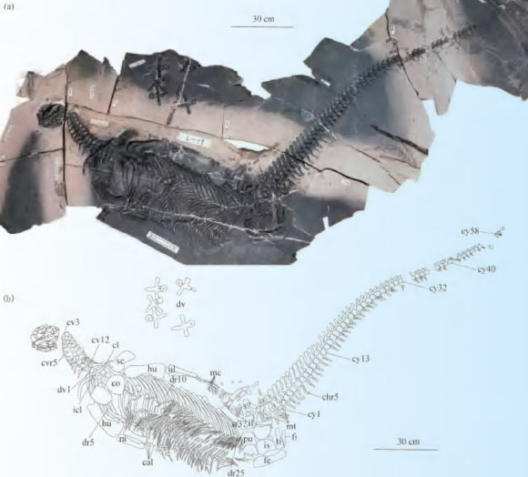
Spoon-Toothed, Short-Headed Marine Reptile Unearthed in China
Un reptile marin à dents de cuillère et à tête courte découvert en Chine
dent Chine Trias Sauropterygia nouvelle espèce autres reptiles squelette
Un nouveau genre et une nouvelle espèce de reptile sauroptérygien ayant vécu pendant la période du Trias, il y a environ 245 millions d'années, ont été identifiés à partir d'un squelette presque complet trouvé en Chine. L'article Un reptile marin à dents de cuillère et à tête courte découvert en Chine est apparu en premier sur Sci.News : Breaking Science News.
20/08/2026 sci-news ⚙ Traduction automatique
Des squelettes vieux de 3 500 ans remettent en question ce que les scientifiques pensaient savoir sur la syphilis
Viêt Nam découverte squelette
Des vestiges anciens du Vietnam suggèrent que des maladies comme le pian pourraient avoir été transmises de la mère à l'enfant il y a des milliers d'années, une forme de transmission souvent supposée indiquer la syphilis. Cette découverte pourrait bouleverser la façon dont les scientifiques interprètent les infections anciennes et compliquer le mystère de longue date des origines de la syphilis.
12/08/2026 sciencedaily-paleo ⚙ Traduction automatique
Ce « crocodile terroriste » de la taille d’un autobus scolaire a mangé des dinosaures. Maintenant c'est de retour
musée fossile Dinosauria Géorgie squelette
Un crocodile massif capable d'attaquer les dinosaures a été ramené à la vie en tant que premier squelette monté scientifiquement précis de Deinosuchus schwimmeri. La réplique de 31 pieds du Tellus Science Museum de Géorgie a été construite à partir d’analyses détaillées de fossiles et de plus de 40 ans de recherches menées par le paléontologue Dr David Schwimmer, en l’honneur duquel l’espèce doit son nom.
05/08/2026 sciencedaily ⚙ Traduction automatique
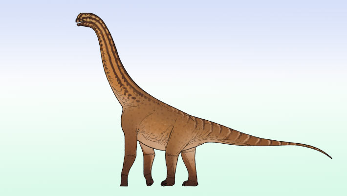
Small Sauropod Dinosaur from Jurassic Period Discovered in India
Un petit dinosaure sauropode du Jurassique découvert en Inde
bassin Inde Jurassique Jurassique moyen Dinosauria partiel squelette
Des paléontologues indiens ont mis au jour un squelette partiel d’un petit dinosaure néosauropode jusqu’alors inconnu dans les roches du Jurassique moyen du bassin de Kachchh, dans le Gujarat. L'article Petit dinosaure sauropode de la période jurassique découvert en Inde apparaît en premier sur Sci.News : Breaking Science News.
21/07/2026 sci-news ⚙ Traduction automatique
1 2 3 4 5 6