←

squelette

Type de vue

162 image(s) · 26 Actualités

Galerie d'images

Plateosaurus engelhardti skeletons "Skelett 2" on exhibit at the museum of the Institute for Geosciences of the Eberhard-Karls-University Tübingen, Germany. "Skelett 2" is a composite from 2 individuals from Trossingen, Germany. Mounts created under the direction of Friedrich von Huene.
Taxons Plateosaurus

Plateosaurus engelhardti skeletons "Skelett 2" on exhibit at the museum of the Institute for Geosciences of the Eberhard-Karls-University Tübingen, Germany. "Skelett 2" is a composite from 2 individuals from Trossingen, Germany. Mounts created under the direction of Friedrich von Huene.

musée Allemagne Plateosauria squelette
Protoceratops andrewsi skeleton at Carnegie Museum of Natural History.
Taxons Protoceratops

Protoceratops andrewsi skeleton at Carnegie Museum of Natural History.

musée Pologne Protoceratops squelette
Reconstructed skeleton of Argentinonectes calafatensis at the Bernardino Rivadavia Natural Sciences Argentine Museum in 2018.
Taxons Argentinonectes

Reconstructed skeleton of Argentinonectes calafatensis at the Bernardino Rivadavia Natural Sciences Argentine Museum in 2018.

musée Argentine Argentinonectes squelette
Ornithischian dinosaur Stenopelix valdensis Meyer, 1857, holotype (GZG 741/2, formerly GPI Gö 741−2), from the Obernkirchen Sandstone (Early
Cretaceous: Berriasian), near Bückeburg, Niedersachsen, Germany. A. Large sandstone slab. B. Interpretative outline drawing of large latex cast (prepared
from large sandstone slab), showing majority of postcranial skeleton in dorsal view. C. Small sandstone slab. D. Interpretative outline drawing of small la−
tex cast (prepared from small sandstone slab), showing sacrum and caudals, pelvic region and partial hindlimbs in ventral view. For clarity elements in and
around the sacral region have not been labelled—these areas are shown in greater detail in Fig. 3. Roman numerals II–IV correspond to respective digits.

Abbreviations: mt, metatarsals; f, femur; ?, unidentified element.
Taxons Stenopelix

Ornithischian dinosaur Stenopelix valdensis Meyer, 1857, holotype (GZG 741/2, formerly GPI Gö 741−2), from the Obernkirchen Sandstone (Early Cretaceous: Berriasian), near Bückeburg, Niedersachsen, Germany. A. Large sandstone slab. B. Interpretative outline drawing of large latex cast (prepared from large sandstone slab), showing majority of postcranial skeleton in dorsal view. C. Small sandstone slab. D. Interpretative outline drawing of small la− tex cast (prepared from small sandstone slab), showing sacrum and caudals, pelvic region and partial hindlimbs in ventral view. For clarity elements in and around the sacral region have not been labelled—these areas are shown in greater detail in Fig. 3. Roman numerals II–IV correspond to respective digits. Abbreviations: mt, metatarsals; f, femur; ?, unidentified element.

dessin Allemagne Berriasien Crétacé +8
Diagnostic skeletal elements of the type specimen of Franconiasaurus brevispinus gen. et sp. nov. (BT 011224.00). (A) main slab showing partly articulated skeleton in dorsal view. Posterior part of right mandibular ramus in (B) lateral and (C) dorsal aspect; anterior cervical vertebra (D) in articular and (E) lateral view; (F) posterior cervical vertebra in lateral view; (G) cervical rib in dorsolateral view; (H) dorsal vertebra in posterior view; (I) left scapula in medial view; (J) right coracoid in supposed dorsal view; (K) left humerus in dorsal view; (L) left femur with adjacent epipodials in dorsal view; and (M) left ilium in lateral view.
Taxons Franconiasaurus

Diagnostic skeletal elements of the type specimen of Franconiasaurus brevispinus gen. et sp. nov. (BT 011224.00). (A) main slab showing partly articulated skeleton in dorsal view. Posterior part of right mandibular ramus in (B) lateral and (C) dorsal aspect; anterior cervical vertebra (D) in articular and (E) lateral view; (F) posterior cervical vertebra in lateral view; (G) cervical rib in dorsolateral view; (H) dorsal vertebra in posterior view; (I) left scapula in medial view; (J) right coracoid in supposed dorsal view; (K) left humerus in dorsal view; (L) left femur with adjacent epipodials in dorsal view; and (M) left ilium in lateral view.

humérus vertèbre spécimen Franconiasaurus +1
Incomplete skeleton of Camptosaurus prestwichii, now referred to as Cumnoria, Oxford University Museum of Natural History.
Taxons Cumnoria

Incomplete skeleton of Camptosaurus prestwichii, now referred to as Cumnoria, Oxford University Museum of Natural History.

musée Camptosaurus Cumnoria squelette
Yueosaurus tiantaiensis, reconstructed skeletons, Zhejiang Geological Museum
Taxons Yueosaurus

Yueosaurus tiantaiensis, reconstructed skeletons, Zhejiang Geological Museum

musée Yueosaurus squelette
Schematic drawing of MUPA-ATZ0103, postcranial skeleton of the paratype of Paludidraco multidentatus.
Taxons Paludidraco

Schematic drawing of MUPA-ATZ0103, postcranial skeleton of the paratype of Paludidraco multidentatus.

dessin Paludidraco squelette
The incomplete skeleton of Cetiosauriscus stewarti (BMNH R3078) mounted prior to display in around
1903. Photographed in the BMNH, and reproduced courtesy of the Leeds family, with layout by J. J. Liston. Note

the similarity of this photograph to the drawing of the skeleton in Woodward (1905); previously figured in Anon. (1924) and Naish & Martill (2008).[1][2]
Taxons Cetiosauriscus

The incomplete skeleton of Cetiosauriscus stewarti (BMNH R3078) mounted prior to display in around 1903. Photographed in the BMNH, and reproduced courtesy of the Leeds family, with layout by J. J. Liston. Note the similarity of this photograph to the drawing of the skeleton in Woodward (1905); previously figured in Anon. (1924) and Naish & Martill (2008).[1][2]

dessin Cetiosauriscus squelette
A–B, skull in dorsal view; C–E, postcranial skeleton; in left dorsolateral (C) and left lateral (D–E) views. In line drawings (B, E) dark grey tone indicates damage and light grey tone indicates the palate. Abbreviations: ca, caudal vertebra [number following indicates order in preserved series]; ce, cervical vertebra; d, dorsal vertebra; depr, depression; ecto, ectopterygoid; epip, epipterygoid; exp, expanded neural spine apex; fr, frontal; jug, jugal; l., left [followed by name of element]; mx, maxilla; p, ‘pectoral’ vertebra; par, parietal; pmx, premaxilla; po, postorbital; pofr, postfrontal; prfr, prefrontal; qua, quadrate; r., right [followed by name of element]; s, sacral vertebra; sq, squamosal; unexp, unexpanded neural spine apex. Scale bars equal 50 mm (A–B), 20 mm (C), and 200 mm (D–E).
Taxons Avalonnectes

A–B, skull in dorsal view; C–E, postcranial skeleton; in left dorsolateral (C) and left lateral (D–E) views. In line drawings (B, E) dark grey tone indicates damage and light grey tone indicates the palate. Abbreviations: ca, caudal vertebra [number following indicates order in preserved series]; ce, cervical vertebra; d, dorsal vertebra; depr, depression; ecto, ectopterygoid; epip, epipterygoid; exp, expanded neural spine apex; fr, frontal; jug, jugal; l., left [followed by name of element]; mx, maxilla; p, ‘pectoral’ vertebra; par, parietal; pmx, premaxilla; po, postorbital; pofr, postfrontal; prfr, prefrontal; qua, quadrate; r., right [followed by name of element]; s, sacral vertebra; sq, squamosal; unexp, unexpanded neural spine apex. Scale bars equal 50 mm (A–B), 20 mm (C), and 200 mm (D–E).

écaille vertèbre dessin Avalonnectes +2
Fossil skeleton of Gargoyleosaurus parkpinorum taken in 2007 at the Denver Museum of Nature and Science.
Taxons Gargoyleosaurus

Fossil skeleton of Gargoyleosaurus parkpinorum taken in 2007 at the Denver Museum of Nature and Science.

musée Denver fossile Gargoyleosaurus +1
Hypnovenator matsubaraetoheorum gen. et sp. nov. Blocks including the forelimb, knee (A), and heel (B). (C) Reconstructed skeleton. Red and yellow colors show the confirmed and probable positions of the recovered elements, respectively (Courtesy of Genya Masukawa). (D) Left manus in medial view. (E) Left manual phalanx I-1 in dorsal view. (F) Removed fragmentary left manual phalanx II-3 (manual ungual phalanx II) for preparing the left manus. (G) Cross-section of the bent right ankle. (H) Left metatarsus in posterior view. Abbreviations: as, astragalus; dp, depression; fe, femur; fi, fibula; hu, humerus; mc I, metacarpal I; mc II, metacarpal II; mc III, metacarpal III; mp I-1, manual phalanx I-1; mp I-2, manual phalanx I-2 (manual ungual phalanx I); mp II-1, manual phalanx II-1; mp II-3, manual phalanx II-3 (manual ungual phalanx II); mp III-3, manual phalanx III-3; mp III-4, manual phalanx III-4 (manual ungual phalanx III); mr, medial ridge; mt II, metatarsal II; mt III, metatarsal III; mt IV, metatarsal IV; mt V, metatarsal V; ra, radius; ti, tibia; ul, ulna. Almost all elements are from the left side. Abbreviations for elements from the right side added ‘(r)’ at the end. This figure was created using Adobe Photoshop 25.5.1 and Adobe Illustrator 28.3 (https://www.adobe.com/).
Taxons Hypnovenator

Hypnovenator matsubaraetoheorum gen. et sp. nov. Blocks including the forelimb, knee (A), and heel (B). (C) Reconstructed skeleton. Red and yellow colors show the confirmed and probable positions of the recovered elements, respectively (Courtesy of Genya Masukawa). (D) Left manus in medial view. (E) Left manual phalanx I-1 in dorsal view. (F) Removed fragmentary left manual phalanx II-3 (manual ungual phalanx II) for preparing the left manus. (G) Cross-section of the bent right ankle. (H) Left metatarsus in posterior view. Abbreviations: as, astragalus; dp, depression; fe, femur; fi, fibula; hu, humerus; mc I, metacarpal I; mc II, metacarpal II; mc III, metacarpal III; mp I-1, manual phalanx I-1; mp I-2, manual phalanx I-2 (manual ungual phalanx I); mp II-1, manual phalanx II-1; mp II-3, manual phalanx II-3 (manual ungual phalanx II); mp III-3, manual phalanx III-3; mp III-4, manual phalanx III-4 (manual ungual phalanx III); mr, medial ridge; mt II, metatarsal II; mt III, metatarsal III; mt IV, metatarsal IV; mt V, metatarsal V; ra, radius; ti, tibia; ul, ulna. Almost all elements are from the left side. Abbreviations for elements from the right side added ‘(r)’ at the end. This figure was created using Adobe Photoshop 25.5.1 and Adobe Illustrator 28.3 (https://www.adobe.com/).

humérus Hypnovenator squelette
Partial skeleton of the heterodontosaurid Tianyulong confuciusi from the Lower Cretaceous Jehol Group of China. Partial skeleton mainly in right lateral view (IVPP V17090). Enlargements of subsequent figures are shown in red. Scale bar equals 10 cm.
Taxons Tianyulong

Partial skeleton of the heterodontosaurid Tianyulong confuciusi from the Lower Cretaceous Jehol Group of China. Partial skeleton mainly in right lateral view (IVPP V17090). Enlargements of subsequent figures are shown in red. Scale bar equals 10 cm.

écaille Chine Crétacé Heterodontosauridae +3
Reconstructed Thalassodromeus sethi skeleton (the postcranium is hypothetical) with Anhanguera behind, National Museum of Nature and Science, Tokyo.
Taxons Thalassodromeus

Reconstructed Thalassodromeus sethi skeleton (the postcranium is hypothetical) with Anhanguera behind, National Museum of Nature and Science, Tokyo.

musée Anhanguera Thalassodromeus squelette
Eustreptospondylus skeleton
Taxons Eustreptospondylus

Eustreptospondylus skeleton

Eustreptospondylus squelette
A photograph of the skeleton of Jianchangosaurus yixianensis gen. et sp. nov. (41HIII-0308A) (A) and line drawing (B).

Abbreviations: at, atlas; ax, axis; cav, caudal vertebra; cv, cervical vertebra; co, coracoid; dc, distal carpal; dv, dorsal vertebra; l., left; f, femur; fi, fibula; fur, furcula; hu, humerus; il, ilium; is, ischium; pu, pubis; ra, radius; sc, scapula; ti, tibia; ul, ulna; mc, metacarpal; mt, metatarsal; mxf, maxillary fenestra; pat, proatlas; r., right; sr, sacral rib; sv, sacral vertebra; ung, ungual. All elements of the skeleton are preserved except the distal half of the caudal vertebrae. Dashed lines of metatarsals indicate areas that have been reconstructed. The middle portion of the neck, from the fourth to ninth cervical vertebrae, and the pedal phalanges have been repositioned. The rest of elements of this specimen are in the original position.
Taxons Jianchangosaurus

A photograph of the skeleton of Jianchangosaurus yixianensis gen. et sp. nov. (41HIII-0308A) (A) and line drawing (B). Abbreviations: at, atlas; ax, axis; cav, caudal vertebra; cv, cervical vertebra; co, coracoid; dc, distal carpal; dv, dorsal vertebra; l., left; f, femur; fi, fibula; fur, furcula; hu, humerus; il, ilium; is, ischium; pu, pubis; ra, radius; sc, scapula; ti, tibia; ul, ulna; mc, metacarpal; mt, metatarsal; mxf, maxillary fenestra; pat, proatlas; r., right; sr, sacral rib; sv, sacral vertebra; ung, ungual. All elements of the skeleton are preserved except the distal half of the caudal vertebrae. Dashed lines of metatarsals indicate areas that have been reconstructed. The middle portion of the neck, from the fourth to ninth cervical vertebrae, and the pedal phalanges have been repositioned. The rest of elements of this specimen are in the original position.

humérus vertèbre dessin spécimen +2
1 2 3 4 5 6 7 8 9 10 11

Actualités

Un fossile remarquable révèle le régime alimentaire d'un ichtyosaure du Jurassique supérieur
Allemagne Jurassique Jurassique supérieur fossile spécimen Aegirosaurus Ichthyosauria Jabalisaurus squelette
Les scientifiques étudiant l’ichtyosaure Jabalisaurus tethyensis, récemment décrit, ont acquis un aperçu remarquable de son régime alimentaire. Un fossile magnifiquement conservé du sud de l'Allemagne contient des preuves des derniers repas de l'animal. De plus, le spécimen préserve des détails importants du squelette et des contours du corps. Le fossile, numéro de spécimen JME-SOS-08369, était auparavant attribué à Aegirosaurus. Cependant, les chercheurs
24/08/2026 everythingdinosaur ⚙ Traduction automatique
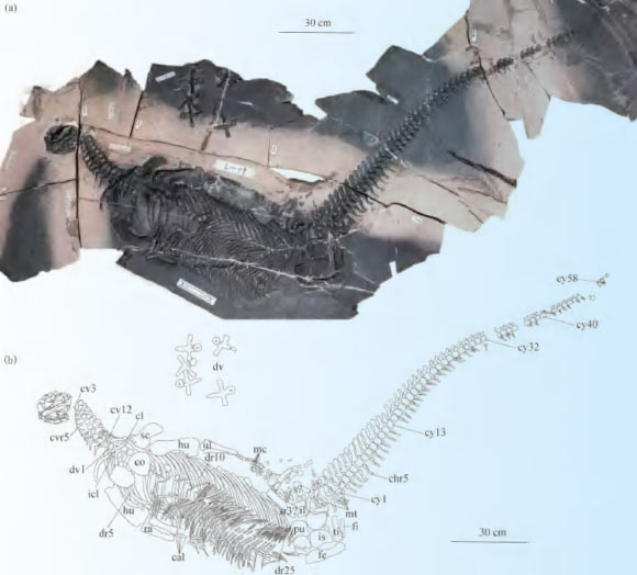
Spoon-Toothed, Short-Headed Marine Reptile Unearthed in China
Un reptile marin à dents de cuillère et à tête courte découvert en Chine
dent Chine Trias Sauropterygia nouvelle espèce autres reptiles squelette
Un nouveau genre et une nouvelle espèce de reptile sauroptérygien ayant vécu pendant la période du Trias, il y a environ 245 millions d'années, ont été identifiés à partir d'un squelette presque complet trouvé en Chine. L'article Un reptile marin à dents de cuillère et à tête courte découvert en Chine est apparu en premier sur Sci.News : Breaking Science News.
20/08/2026 sci-news ⚙ Traduction automatique
Des squelettes vieux de 3 500 ans remettent en question ce que les scientifiques pensaient savoir sur la syphilis
Viêt Nam découverte squelette
Des vestiges anciens du Vietnam suggèrent que des maladies comme le pian pourraient avoir été transmises de la mère à l'enfant il y a des milliers d'années, une forme de transmission souvent supposée indiquer la syphilis. Cette découverte pourrait bouleverser la façon dont les scientifiques interprètent les infections anciennes et compliquer le mystère de longue date des origines de la syphilis.
12/08/2026 sciencedaily-paleo ⚙ Traduction automatique
Ce « crocodile terroriste » de la taille d’un autobus scolaire a mangé des dinosaures. Maintenant c'est de retour
musée fossile Dinosauria Géorgie squelette
Un crocodile massif capable d'attaquer les dinosaures a été ramené à la vie en tant que premier squelette monté scientifiquement précis de Deinosuchus schwimmeri. La réplique de 31 pieds du Tellus Science Museum de Géorgie a été construite à partir d’analyses détaillées de fossiles et de plus de 40 ans de recherches menées par le paléontologue Dr David Schwimmer, en l’honneur duquel l’espèce doit son nom.
05/08/2026 sciencedaily ⚙ Traduction automatique
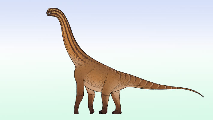
Small Sauropod Dinosaur from Jurassic Period Discovered in India
Un petit dinosaure sauropode du Jurassique découvert en Inde
bassin Inde Jurassique Jurassique moyen Dinosauria partiel squelette
Des paléontologues indiens ont mis au jour un squelette partiel d’un petit dinosaure néosauropode jusqu’alors inconnu dans les roches du Jurassique moyen du bassin de Kachchh, dans le Gujarat. L'article Petit dinosaure sauropode de la période jurassique découvert en Inde apparaît en premier sur Sci.News : Breaking Science News.
21/07/2026 sci-news ⚙ Traduction automatique
1 2 3 4 5 6