←

skull

View type

215 image(s) · 30 News

Image gallery

Hualianceratops holotype IVVP V18641, Skull reconstruction
Taxa Hualianceratops

Hualianceratops holotype IVVP V18641, Skull reconstruction

holotype Hualianceratops skull
Figure 2.
Holotype of Liaodactylus primus gen. et sp. nov. (PMOL-AP00031): photographs and line drawings of the nearly complete skull with mandibles in left lateral (a,b), and palatal (c,d) views. aa, atlas-axis complex; an, angular; arf, articular facet; bo, basioccipital; bs, basisphenoid; co, coronoid; d, dentary; ec, ectopterygoid; f, frontal; j, jugal; m, maxilla; naof, nasoantorbital fenestra; p, parietal; pal, palatine; pat, proatlas; pecf, pterygo-ectopterygoid fenestra; pm, premaxilla; pof, postfrontal; pra, prearticular; pt, pterygoid; q, quadrate; qj, quadratojugal; sa, surangular; sof, suborbital fenestra; spl, splenial; sq, squamosal; stf, subtemporal fenestra.
Taxa Liaodactylus

Figure 2. Holotype of Liaodactylus primus gen. et sp. nov. (PMOL-AP00031): photographs and line drawings of the nearly complete skull with mandibles in left lateral (a,b), and palatal (c,d) views. aa, atlas-axis complex; an, angular; arf, articular facet; bo, basioccipital; bs, basisphenoid; co, coronoid; d, dentary; ec, ectopterygoid; f, frontal; j, jugal; m, maxilla; naof, nasoantorbital fenestra; p, parietal; pal, palatine; pat, proatlas; pecf, pterygo-ectopterygoid fenestra; pm, premaxilla; pof, postfrontal; pra, prearticular; pt, pterygoid; q, quadrate; qj, quadratojugal; sa, surangular; sof, suborbital fenestra; spl, splenial; sq, squamosal; stf, subtemporal fenestra.

drawing holotype Liaodactylus skull
The skull of Zupaysaurus rougieri (A) in right lateral view. Scale bar equals 5 cm. Cropped from Figure 2 in the source.
anfe - antorbital fenestra
emf - external mandibular fenestra
itf - infratemporal fenestra
or - orbit
Taxa Zupaysaurus

The skull of Zupaysaurus rougieri (A) in right lateral view. Scale bar equals 5 cm. Cropped from Figure 2 in the source. anfe - antorbital fenestra emf - external mandibular fenestra itf - infratemporal fenestra or - orbit

scale Zupaysaurus skull
Skeletal elements of Bajadasaurus pronuspinax gen. et sp. nov (MMCh-PV 75). (A–C) Skull roof and braincase in posterior (A), left lateral (B) and right lateral (C) views. (D,E) Left lower jaw in dorsal (D) and medial (E) views. F, Dentaries in anterior view. (G) Pterygoids in ventral view. (H) Left maxilla in medial view. (I) Left lacrimal in lateral view. (J) Left quadratojugal in lateral view. (K,L) Right quadrate in medial (K) and posterior (L) views. (M) Proatlases in dorsal view. (N) Atlantal neurapophyses in anterior view. (O,P), Axis in left lateral (O) and anterior (P) views. (Q,R) Fifth cervical vertebra in left lateral (Q) and anterior (R) views. an, angular; ar, articular; bo, basioccipital; bt, basal tubera; btp, basipterygoid process; ch, ‘chin’ of dentary; cn, cranial nerve; d, dentary; di, diapophysis; f, frontal; fm, foramen magnum; fo, fenestra ovalis; ls, laterosphenoid; met, metotic foramen; mp, medial process; nc, neural canal; ns, neural spine; os, orbitosphenoid; p, parietal; pfo, pneumatic fossa; po, postorbital; pocdf, postzygapophyseal centrodiapophyseal fossa; podl, postzygodiapophyseal lamina; popr, paraoccipital process; poz, postzygapophysis; pra, prearticular; pre, prezygapophysis; prsl, prespinal lamina; ptf, postemporal fenestra; qf, quadrate fossa; rm, replacement maxillary tooth; sa, surangular; sp, splenial; sq, squamosal; stf, supratemporal fenestra; vk, ventral keel; vp, ventral process.
Taxa Bajadasaurus

Skeletal elements of Bajadasaurus pronuspinax gen. et sp. nov (MMCh-PV 75). (A–C) Skull roof and braincase in posterior (A), left lateral (B) and right lateral (C) views. (D,E) Left lower jaw in dorsal (D) and medial (E) views. F, Dentaries in anterior view. (G) Pterygoids in ventral view. (H) Left maxilla in medial view. (I) Left lacrimal in lateral view. (J) Left quadratojugal in lateral view. (K,L) Right quadrate in medial (K) and posterior (L) views. (M) Proatlases in dorsal view. (N) Atlantal neurapophyses in anterior view. (O,P), Axis in left lateral (O) and anterior (P) views. (Q,R) Fifth cervical vertebra in left lateral (Q) and anterior (R) views. an, angular; ar, articular; bo, basioccipital; bt, basal tubera; btp, basipterygoid process; ch, ‘chin’ of dentary; cn, cranial nerve; d, dentary; di, diapophysis; f, frontal; fm, foramen magnum; fo, fenestra ovalis; ls, laterosphenoid; met, metotic foramen; mp, medial process; nc, neural canal; ns, neural spine; os, orbitosphenoid; p, parietal; pfo, pneumatic fossa; po, postorbital; pocdf, postzygapophyseal centrodiapophyseal fossa; podl, postzygodiapophyseal lamina; popr, paraoccipital process; poz, postzygapophysis; pra, prearticular; pre, prezygapophysis; prsl, prespinal lamina; ptf, postemporal fenestra; qf, quadrate fossa; rm, replacement maxillary tooth; sa, surangular; sp, splenial; sq, squamosal; stf, supratemporal fenestra; vk, ventral keel; vp, ventral process.

tooth vertebra Bajadasaurus skull
The skull of Aquilops in three versions: the actual holotype fossil down below, a reconstruction of the distorted skull with the missing bits sculpted in by preparator and reptile-reconstructor extraordinaire Kyle Davies (see more of his work here), and a reconstruction of the skull as it might have looked before it went through the ravages of taphonomy.
Taxa Aquilops

The skull of Aquilops in three versions: the actual holotype fossil down below, a reconstruction of the distorted skull with the missing bits sculpted in by preparator and reptile-reconstructor extraordinaire Kyle Davies (see more of his work here), and a reconstruction of the skull as it might have looked before it went through the ravages of taphonomy.

fossil holotype Aquilops skull
Figure 1: Holotype and skeletal reconstruction of Mosaiceratops azumai, gen. et sp. nov (ZMNH M8856). (a) photograph and line drawing of ZMNH M8856; (b) skeletal reconstruction showing preserved elements in white. Scale bar 10 cm. Abbreviations: a, astragalus; boc, basioccipital; c, calcaneum; cav, caudal vertebra; ch, chevron; cv, cervical vertebra; dr, dorsal rib; dv, dorsal vertebra; f, frontal; fem, femur; fl, fibula; h, humerus; il, ilium; is, ischium; L, left; mt, metatarsal; ph, phalanx/phalanges; po, postorbital; R, right; sk, skull; sq, squamosal; t, tibia; td, tendon; ?, undiagnostic remains.
Taxa Mosaiceratops

Figure 1: Holotype and skeletal reconstruction of Mosaiceratops azumai, gen. et sp. nov (ZMNH M8856). (a) photograph and line drawing of ZMNH M8856; (b) skeletal reconstruction showing preserved elements in white. Scale bar 10 cm. Abbreviations: a, astragalus; boc, basioccipital; c, calcaneum; cav, caudal vertebra; ch, chevron; cv, cervical vertebra; dr, dorsal rib; dv, dorsal vertebra; f, frontal; fem, femur; fl, fibula; h, humerus; il, ilium; is, ischium; L, left; mt, metatarsal; ph, phalanx/phalanges; po, postorbital; R, right; sk, skull; sq, squamosal; t, tibia; td, tendon; ?, undiagnostic remains.

humerus scale vertebra drawing +3
Skull of Eternauta patagonica (photograph and diagram in right view)
Taxa Eternauta

Skull of Eternauta patagonica (photograph and diagram in right view)

Eternauta skull
Skeletal diagram featuring the optimal remains of the holotype of Graciliceratops mongoliensis: ZPAL MgD-I/156.[1] Found in the Bayan Shireh Formation, in the original description the remains were referred to the genus Microceratops (now obsolete).[2] However, Sereno in 2000 noted that there was no base for this referral, then, he created a new genus and species for this specimen.[1] The holotype is very fragmented (specially the skull), consisting of:[2]
Fragmented skull; 4 cervical, 12 dorsal and 7 sacral vertebrae; right scapula; proximal end of left scapula; left coracoid; right humerus, radius and fragmentary ulna; proximal and distal end of left humerus; proximal fragments of both pubis; fragments of both illium and fragment of right ischium; right femur, tibia and nearly complete pes; distal part of left tibia, fragmentary left pes; tarsals and isolated ribs.[2]

The sacral vertebrae are not fused, an indicator of the immaturity of this specimen; the estimated adult size is about 2 meters long or similar to Protoceratops.[2][1] Right quadratojugal, quadrate and fragmentary jugal were reversed in order to get an optimal view.
Taxa Graciliceratops

Skeletal diagram featuring the optimal remains of the holotype of Graciliceratops mongoliensis: ZPAL MgD-I/156.[1] Found in the Bayan Shireh Formation, in the original description the remains were referred to the genus Microceratops (now obsolete).[2] However, Sereno in 2000 noted that there was no base for this referral, then, he created a new genus and species for this specimen.[1] The holotype is very fragmented (specially the skull), consisting of:[2] Fragmented skull; 4 cervical, 12 dorsal and 7 sacral vertebrae; right scapula; proximal end of left scapula; left coracoid; right humerus, radius and fragmentary ulna; proximal and distal end of left humerus; proximal fragments of both pubis; fragments of both illium and fragment of right ischium; right femur, tibia and nearly complete pes; distal part of left tibia, fragmentary left pes; tarsals and isolated ribs.[2] The sacral vertebrae are not fused, an indicator of the immaturity of this specimen; the estimated adult size is about 2 meters long or similar to Protoceratops.[2][1] Right quadratojugal, quadrate and fragmentary jugal were reversed in order to get an optimal view.

humerus description holotype specimen +5
Zhejiangopterus linhaiensis, skull (cast), Zhejiang Geological Museum
Taxa Zhejiangopterus

Zhejiangopterus linhaiensis, skull (cast), Zhejiang Geological Museum

museum cast Zhejiangopterus skull
Smitanosaurus agilis skull fragment and neck.
Taxa Smitanosaurus

Smitanosaurus agilis skull fragment and neck.

Smitanosaurus skull
The theropod skull displays the distinctive features of this apex predator, including a long, robust snout, conical teeth, and strong jaw muscles adapted for gripping and tearing prey.
Taxa Rajasaurus

The theropod skull displays the distinctive features of this apex predator, including a long, robust snout, conical teeth, and strong jaw muscles adapted for gripping and tearing prey.

predator prey Rajasaurus skull
Reconstruction of the skull of Tlatolophus galorum by sculptor Samuel Nieves Tlapaya (El Alebrije Prehistórico). Displayed in the lobby of the hosting hotel of the 12th Latin American Congress of Paleontology, held in Puebla, Mexico, during the first week of March 2025.
Taxa Tlatolophus

Reconstruction of the skull of Tlatolophus galorum by sculptor Samuel Nieves Tlapaya (El Alebrije Prehistórico). Displayed in the lobby of the hosting hotel of the 12th Latin American Congress of Paleontology, held in Puebla, Mexico, during the first week of March 2025.

Mexico Tlatolophus skull
Skull of Caiuajara dobruskii gen. et sp. nov. (holotype, CP.V 1449) with the shape of an adult individual.

Scale bar equals 50: d, dentary; dcr, dentary crest; dep, depression; exp, ventral expansion of the premaxilla; f, frontal; fcr, frontal crest; fo, foraminae; m, maxilla; oc, occipital condyle; op, opisthotic; p, parietal; pm, premaxilla; pmcr, premaxillary crest; q, quadrate; soc, supraoccipital. The quadrate is inverted.
Taxa Caiuajara

Skull of Caiuajara dobruskii gen. et sp. nov. (holotype, CP.V 1449) with the shape of an adult individual. Scale bar equals 50: d, dentary; dcr, dentary crest; dep, depression; exp, ventral expansion of the premaxilla; f, frontal; fcr, frontal crest; fo, foraminae; m, maxilla; oc, occipital condyle; op, opisthotic; p, parietal; pm, premaxilla; pmcr, premaxillary crest; q, quadrate; soc, supraoccipital. The quadrate is inverted.

crest scale holotype Caiuajara +1
Minqaria bata skull reconstruction with known elements shown in white.
Taxa Minqaria

Minqaria bata skull reconstruction with known elements shown in white.

Minqaria skull
Caupedactylus ybaka holotype skull (MN 4726-V) in D, E, left lateral view.
Taxa Caupedactylus

Caupedactylus ybaka holotype skull (MN 4726-V) in D, E, left lateral view.

holotype Caupedactylus skull
A–B, skull in dorsal view; C–E, postcranial skeleton; in left dorsolateral (C) and left lateral (D–E) views. In line drawings (B, E) dark grey tone indicates damage and light grey tone indicates the palate. Abbreviations: ca, caudal vertebra [number following indicates order in preserved series]; ce, cervical vertebra; d, dorsal vertebra; depr, depression; ecto, ectopterygoid; epip, epipterygoid; exp, expanded neural spine apex; fr, frontal; jug, jugal; l., left [followed by name of element]; mx, maxilla; p, ‘pectoral’ vertebra; par, parietal; pmx, premaxilla; po, postorbital; pofr, postfrontal; prfr, prefrontal; qua, quadrate; r., right [followed by name of element]; s, sacral vertebra; sq, squamosal; unexp, unexpanded neural spine apex. Scale bars equal 50 mm (A–B), 20 mm (C), and 200 mm (D–E).
Taxa Avalonnectes

A–B, skull in dorsal view; C–E, postcranial skeleton; in left dorsolateral (C) and left lateral (D–E) views. In line drawings (B, E) dark grey tone indicates damage and light grey tone indicates the palate. Abbreviations: ca, caudal vertebra [number following indicates order in preserved series]; ce, cervical vertebra; d, dorsal vertebra; depr, depression; ecto, ectopterygoid; epip, epipterygoid; exp, expanded neural spine apex; fr, frontal; jug, jugal; l., left [followed by name of element]; mx, maxilla; p, ‘pectoral’ vertebra; par, parietal; pmx, premaxilla; po, postorbital; pofr, postfrontal; prfr, prefrontal; qua, quadrate; r., right [followed by name of element]; s, sacral vertebra; sq, squamosal; unexp, unexpanded neural spine apex. Scale bars equal 50 mm (A–B), 20 mm (C), and 200 mm (D–E).

scale vertebra drawing Avalonnectes +2
1 2 3 4 5 6 7 8 9 10 11 12 13 14

News

Son crâne mesure 2 mètres et compte encore 130 dents : découvrez ce redoutable prédateur du Jurassique
tooth predator book Jurassic Pliosaurus discovery skull
Une découverte paléontologique majeure vient de secouer la communauté scientifique mondiale. Un crâne gigantesque de pliosaure, reptile marin datant de 150 millions d'années, a récemment été inscrit au Guinness Book des records. Cette relique exceptionnellement préservée nous offre une fenêtre...
04/10/2026 futura-terre
Paleontologists Identify New Species of Glyptodont
museum Denmark new species skull
An incomplete fossilized skull that has sat in the collections of the Natural History Museum of Denmark since the 19th century has turned out to represent a previously unrecognized species of glyptodont (genus Glyptotherium) that once roamed South America. The post Paleontologists Identify New Species of Glyptodont appeared first on Sci.News: Breaking Science News.
28/08/2026 sci-news
Remarkable New Museum für Naturkunde Berlin Galleries Open to Visitors
museum skull
Two remarkable Museum für Naturkunde Berlin galleries are being opened to visitors for the first time. The new "Mammal Skulls" and "Deer and Horned Animals" galleries will welcome members of the public during the Long Night of Museums event on Saturday 29th of August. Until now, these collections have been reserved exclusively for researchers. Their
28/08/2026 everythingdinosaur
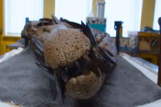
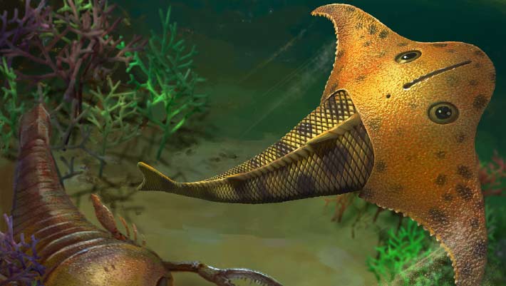
Meet Bataspis crux, Jawless Fish that Cruised Devonian Seas with Batwing Head
jaw China Devonian Late Devonian fish new species skull
Paleontologists in China have identified a new species of armored, jawless fish from rocks around 419 million years old whose skull was shaped like the wing of a bat and whose unusual head made it an efficient glider through Devonian seas. The post Meet Bataspis crux, Jawless Fish that Cruised Devonian Seas with Batwing Head appeared first on Sci.News: Breaking Science News.
18/08/2026 sci-news
A 30-year-old fossil was hiding bones from a mysterious ancient sea monster
bone jaw Cretaceous fossil Mosasaurus new species skull
Fossils collected in Osaka more than 30 years ago have revealed a surprise hiding in plain sight: four previously unrecognized bones from a giant Cretaceous mosasaur. Among them is the first confirmed premaxilla—the frontmost bone of the upper jaw—ever identified in a Japanese mosasaur fossil. Even more intriguing, parts of the skull show unusual anatomical features that don’t match known mosasaurs, raising the possibility that the remains belong to an entirely new species.
15/08/2026 sciencedaily
1 2 3 4 5 6