dent
Partie anatomique55 image(s) · 28 Actualités
Galerie d'images


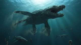
![Reconstruction of the terrestrial paleoenvironmental setting of the Sao Khua Formation by Renata Cunha.
In the center, a generalized spinosaurid feeds on a sauropod. This trophic relationship is hypothesized based on isolated tooth crowns found in association with a sauropod skeleton [67]. In the background, a small pack of the ornithomimosaur theropod Kinnareemimus. Both sauropods and ornithomimosaurs (as part of the “herbivorous” theropods) were found to be positively associated with terrestrial paleoenvironments by Butler and Barrett [15].
(cropped from File:Spinosaurid and Kinnareemimus.PNG)](https://upload.wikimedia.org/wikipedia/commons/thumb/6/62/Kinnareemimus_pack.png/330px-Kinnareemimus_pack.png)


Actualités
05/07/2026
sciencedaily
⚙ Traduction automatique

29/06/2026
sci-news
⚙ Traduction automatique
os
dent
vertèbre
chasse
musée
Jurassique
fossile
Ichthyosauria
Ichthyosaurus
Pliosaurus
comportement
17/06/2026
futura-terre

15/06/2026
sci-news
⚙ Traduction automatique