←

dent

Partie anatomique

56 image(s) · 40 Actualités

Galerie d'images

Fig. 2. Side view of crown of tooth of Cardiodon rugulosus.
Fig. 3. Fore end of the same tooth.
Fig. 4. Hind end of the crown of another tooth of Cardiodon rugulosus.
Fig. 5. Magnified view of markings on the surface of the enamel of the same tooth.
All the figures are of the natural size.

2-5 are from the Forest Marble of Bradford, Wilts. In the Collection of Channing Pearce, Esq., of that town.
Taxons Cardiodon

Fig. 2. Side view of crown of tooth of Cardiodon rugulosus. Fig. 3. Fore end of the same tooth. Fig. 4. Hind end of the crown of another tooth of Cardiodon rugulosus. Fig. 5. Magnified view of markings on the surface of the enamel of the same tooth. All the figures are of the natural size. 2-5 are from the Forest Marble of Bradford, Wilts. In the Collection of Channing Pearce, Esq., of that town.

dent Cardiodon
Fig. 1. Type material of Amygdalodon patagonicus Cabrera. 1947. A. B. Tooth, MLP 46-VIII-21-1/13, in lingual (A) and labial (B) view. C. D, E. Cervical vertebra, MLP 46-VIII-21-1/8. in right lateral (C, stereopair), ventral (D) and left lateral (E) view. F, G. Right cervical prezygapophysis, MLP 46-VIII-21-117. in dorsal (F) and medial (G) view. H. Anterior dorsal neural spine, MLP 46-VIII-21-116. in posterior view. I, J, K. Posterior dorsal vertebra, MLP 46-VIII-21-112, lectotype, in right lateral view (I, stereopair), right lateral view, with right wall of neural canal remived (J), and posterior (K) view. L, M. Caudal vertebra, MLP 46-VIII-21-1/3, in left lateral (L) and posterior (M) view. N. Caudal vertebra, MLP 46-VIII-21-1/4, in right lateral view. (O). Proximal fragment of a dorsal rib, MLP 46-VIII-21-1/9, in anterolateral view. Abbreviations: acdl, anterior centrodiapophyseal lamina; cprl, centroprezygapophyseal lamina; g, groove; k, ventral keel; pcdl, posterior centrodiapophyseal lamina; pod, postzygodiapophyseal lamina; poz, postzygapophysis; pp, parapophysis; prdl, prezygodiapophyseal lamina; spol, spinopostzgapophyseal lamina; sprl, spinoprezygapophyseal lamina; tprl, intraprezygapophyseal lamina. Scale bars 1 cm (A, B) and 10 cm (C-0).
Taxons Amygdalodon

Fig. 1. Type material of Amygdalodon patagonicus Cabrera. 1947. A. B. Tooth, MLP 46-VIII-21-1/13, in lingual (A) and labial (B) view. C. D, E. Cervical vertebra, MLP 46-VIII-21-1/8. in right lateral (C, stereopair), ventral (D) and left lateral (E) view. F, G. Right cervical prezygapophysis, MLP 46-VIII-21-117. in dorsal (F) and medial (G) view. H. Anterior dorsal neural spine, MLP 46-VIII-21-116. in posterior view. I, J, K. Posterior dorsal vertebra, MLP 46-VIII-21-112, lectotype, in right lateral view (I, stereopair), right lateral view, with right wall of neural canal remived (J), and posterior (K) view. L, M. Caudal vertebra, MLP 46-VIII-21-1/3, in left lateral (L) and posterior (M) view. N. Caudal vertebra, MLP 46-VIII-21-1/4, in right lateral view. (O). Proximal fragment of a dorsal rib, MLP 46-VIII-21-1/9, in anterolateral view. Abbreviations: acdl, anterior centrodiapophyseal lamina; cprl, centroprezygapophyseal lamina; g, groove; k, ventral keel; pcdl, posterior centrodiapophyseal lamina; pod, postzygodiapophyseal lamina; poz, postzygapophysis; pp, parapophysis; prdl, prezygodiapophyseal lamina; spol, spinopostzgapophyseal lamina; sprl, spinoprezygapophyseal lamina; tprl, intraprezygapophyseal lamina. Scale bars 1 cm (A, B) and 10 cm (C-0).

écaille dent vertèbre Amygdalodon
Selected elements used in the diagnosis of Saltriovenator zanellai n. gen. n. sp.

Right humerus in medial (A), frontal (B) and distal (C) views; (D) left scapula, medial view; (E) right scapular glenoid and coracoid, lateral view; (F) furcula, ventral view; tooth, labial (G) and apical (H) views; (I) left humerus, medial view; right second metacarpal in dorsal (J), lateral (L) and distal (N) views; first phalanx of the right second digit in dorsal (K), lateral (M) and proximal (O) views; (P–T) right third digit in proximal, dorsal and lateral views; (U) right distal tarsal IV, proximal view; third right metatarsal in proximal (V) and frontal (X) views; second right metatarsal, proximal (W) and frontal (Y) views; (Z) reconstructed skeleton showing identified elements (red). Abbreviations as in text, asterisks mark autapomorphic traits. Scale bars: 10 cm in (A)–(E), (I), and (U)–(Y); two cm in (F), and (J)–(T); one cm in (G).
Taxons Saltriovenator

Selected elements used in the diagnosis of Saltriovenator zanellai n. gen. n. sp. Right humerus in medial (A), frontal (B) and distal (C) views; (D) left scapula, medial view; (E) right scapular glenoid and coracoid, lateral view; (F) furcula, ventral view; tooth, labial (G) and apical (H) views; (I) left humerus, medial view; right second metacarpal in dorsal (J), lateral (L) and distal (N) views; first phalanx of the right second digit in dorsal (K), lateral (M) and proximal (O) views; (P–T) right third digit in proximal, dorsal and lateral views; (U) right distal tarsal IV, proximal view; third right metatarsal in proximal (V) and frontal (X) views; second right metatarsal, proximal (W) and frontal (Y) views; (Z) reconstructed skeleton showing identified elements (red). Abbreviations as in text, asterisks mark autapomorphic traits. Scale bars: 10 cm in (A)–(E), (I), and (U)–(Y); two cm in (F), and (J)–(T); one cm in (G).

humérus écaille dent dessin +2
Solitary tooth of a sauropod from Angeac-Charente in France. Based on its morphology, it is not from an unnamed titanosaur like other material from this site, but from an unnamed titanosauriform similar to Ligabuesaurus or Veneosaurus.
Taxons Ligabuesaurus

Solitary tooth of a sauropod from Angeac-Charente in France. Based on its morphology, it is not from an unnamed titanosaur like other material from this site, but from an unnamed titanosauriform similar to Ligabuesaurus or Veneosaurus.

dent France Ligabuesaurus Titanosauria +1
Cast of the holotype tooth (MGUH DK No. 315) of Dromaeosauroides bornholmensis at the Natural History Museum, London.
Taxons Dromaeosauroides

Cast of the holotype tooth (MGUH DK No. 315) of Dromaeosauroides bornholmensis at the Natural History Museum, London.

dent musée moulage holotype +1
Haliskia peterseni gen. et sp. nov., holotype specimen KK F1426. (A) dorsal vertebra, ribs, gastralium, left scapulocoracoid, left and right manual phalanx IV-2, right manual phalanx IV-3, left femur, left tibia, metatarsals and pedal phalanges, (B) mandible, teeth, ceratobranchials, cervical vertebra, rib, gastralium, right syncarpus, right lateral carpal, right pteroid, metacarpals, manual digits, right manual phalanx IV-1, left manual phalanx IV-4, (C) premaxilla with teeth, (D) isolated tooth, (E) isolated tooth, (F) left lateral carpal, (G) left metacarpal IV. Scale bar = 50 mm.
Taxons Haliskia

Haliskia peterseni gen. et sp. nov., holotype specimen KK F1426. (A) dorsal vertebra, ribs, gastralium, left scapulocoracoid, left and right manual phalanx IV-2, right manual phalanx IV-3, left femur, left tibia, metatarsals and pedal phalanges, (B) mandible, teeth, ceratobranchials, cervical vertebra, rib, gastralium, right syncarpus, right lateral carpal, right pteroid, metacarpals, manual digits, right manual phalanx IV-1, left manual phalanx IV-4, (C) premaxilla with teeth, (D) isolated tooth, (E) isolated tooth, (F) left lateral carpal, (G) left metacarpal IV. Scale bar = 50 mm.

écaille dent vertèbre holotype +2
Outline of Irisosaurus yimenensis displaying the preserved material. The most informative elements are figured. (a) Outline; (b) Tooth; (c) Left maxilla; (d) Middle cervical; (e) Posterior cervical; (f) Anterior dorsal; (g) Middle dorsal neural spine; (h) Right scapula; (i) Right humerus; (j) Right ulna; (k) Right manus. Scale bars = 1 m (a); 1 cm (b); 5 cm (c–g); 10 cm (h–k).
Taxons Irisosaurus

Outline of Irisosaurus yimenensis displaying the preserved material. The most informative elements are figured. (a) Outline; (b) Tooth; (c) Left maxilla; (d) Middle cervical; (e) Posterior cervical; (f) Anterior dorsal; (g) Middle dorsal neural spine; (h) Right scapula; (i) Right humerus; (j) Right ulna; (k) Right manus. Scale bars = 1 m (a); 1 cm (b); 5 cm (c–g); 10 cm (h–k).

humérus écaille dent Irisosaurus
Photographs of Melkamter pateko (MPEF-PV 11530), visible in medial views on the main block (1) with inset of the tooth (1B) and counterslab (2);. Rendered CT scan detail images (dashed areas represent visible bone that was too thin to be captured by CT scan) of the cranial fragment (1A), manual metacarpal (1C) and dorsal vertebrae (1D–F). Abbreviations: a = alveoli; if = infratemporal fenestra; j = jugal; l = lacrimal; m = maxilla; o = orbit; naof = Nnasoantorbital fenestra; np = nasal process; pm = premaxilla; po = postorbital; q = quadrate; qj = quadratojugal; s = squamosal; t = tooth; vas = vestigial ascending process.
Taxons Melkamter

Photographs of Melkamter pateko (MPEF-PV 11530), visible in medial views on the main block (1) with inset of the tooth (1B) and counterslab (2);. Rendered CT scan detail images (dashed areas represent visible bone that was too thin to be captured by CT scan) of the cranial fragment (1A), manual metacarpal (1C) and dorsal vertebrae (1D–F). Abbreviations: a = alveoli; if = infratemporal fenestra; j = jugal; l = lacrimal; m = maxilla; o = orbit; naof = Nnasoantorbital fenestra; np = nasal process; pm = premaxilla; po = postorbital; q = quadrate; qj = quadratojugal; s = squamosal; t = tooth; vas = vestigial ascending process.

os dent Melkamter tomographie
Skull in dorsal (a and b, photograph and drawing respectively), and left lateral (c and d, photograph and drawing respectively) views. (e) Premaxillary tooth; (f,g) maxillary teeth (g inverted). amf, anterior maxillary fossa; aof, antorbital fossa; aso, anterior supraorbital; d, dentary; ef, elliptical fossa; f, foramina; fr, frontal; ift, infratemporal fenestra; j, jugal; mx, maxilla; n, nasals; o, orbit; pd, predentary; pdb, postdentary bones; pmx, premaxilla; po, postorbital; pso: posterior supraorbital; prf, prefrontal; qj, quadratojugal; sq, squamosal; stf, supratemporal fenestra. 1–7 denticles. The drawings were processed using Adobe Photoshop CS2 Serial Number: 1045-1412-5685-1654-6343-1431.
Taxons Isaberrysaura

Skull in dorsal (a and b, photograph and drawing respectively), and left lateral (c and d, photograph and drawing respectively) views. (e) Premaxillary tooth; (f,g) maxillary teeth (g inverted). amf, anterior maxillary fossa; aof, antorbital fossa; aso, anterior supraorbital; d, dentary; ef, elliptical fossa; f, foramina; fr, frontal; ift, infratemporal fenestra; j, jugal; mx, maxilla; n, nasals; o, orbit; pd, predentary; pdb, postdentary bones; pmx, premaxilla; po, postorbital; pso: posterior supraorbital; prf, prefrontal; qj, quadratojugal; sq, squamosal; stf, supratemporal fenestra. 1–7 denticles. The drawings were processed using Adobe Photoshop CS2 Serial Number: 1045-1412-5685-1654-6343-1431.

os dent dessin Isaberrysaura +1
Ziapelta sanjuanensis, gen. et sp. nov., (holotype NMMNH P-64484), complete skull.

A, dorsal view; B, ventral view; C, anterior view; D, occipital view; and E, left lateral view. Abbreviations: asca, anterior supraorbital caputegulum; bas, basioccipital; ch, choana; fm, foramen magnum; j, jugal; laca, lacrimal caputegulum; loca, loreal caputegulum; ltf, laterotemporal fenestra; mnca, median nasal caputegulum; nar, external naris; oc, occipital condyle; orb, orbit; pal, palatine; par, parietal; parocc, paroccipital process; pmx, premaxilla; psca, posterior supraorbital caputegulum; pt, pterygoid; q, quadrate; qj, quadratojugal; qjh, quadratojugal horn; snca, supranarial caputegulum; socc, supraoccipital; sqh, squamosal horn; tr, tooth row; v, vomer.
Taxons Ziapelta

Ziapelta sanjuanensis, gen. et sp. nov., (holotype NMMNH P-64484), complete skull. A, dorsal view; B, ventral view; C, anterior view; D, occipital view; and E, left lateral view. Abbreviations: asca, anterior supraorbital caputegulum; bas, basioccipital; ch, choana; fm, foramen magnum; j, jugal; laca, lacrimal caputegulum; loca, loreal caputegulum; ltf, laterotemporal fenestra; mnca, median nasal caputegulum; nar, external naris; oc, occipital condyle; orb, orbit; pal, palatine; par, parietal; parocc, paroccipital process; pmx, premaxilla; psca, posterior supraorbital caputegulum; pt, pterygoid; q, quadrate; qj, quadratojugal; qjh, quadratojugal horn; snca, supranarial caputegulum; socc, supraoccipital; sqh, squamosal horn; tr, tooth row; v, vomer.

dent holotype Ziapelta crâne
DMNH 2021-05-02, holotype left dentary of Ampelognathus coheni, photos and labeled outline drawings in A, lateral; B, medial; C, dorsal; D, ventral; E, anterior; and F, posterior views. Abbreviations: a#, alveolus and numerical position in tooth row; anf, angular facet; cof, coronoid facet; cop, coronoid process; d6, dentary tooth 6 base; ds, dentary symphysis; mc, Meckelian canal; mg, Meckelian groove; pdd, dorsal facet for predentary; pdv, ventral facet for predentary; saf, surangular facet. Gray fill indicates recessed surfaces. Stippling indicates visible sediment. Cross-hatching indicates broken bone surface. Scale bars equal 5 mm.
Taxons Ampelognathus

DMNH 2021-05-02, holotype left dentary of Ampelognathus coheni, photos and labeled outline drawings in A, lateral; B, medial; C, dorsal; D, ventral; E, anterior; and F, posterior views. Abbreviations: a#, alveolus and numerical position in tooth row; anf, angular facet; cof, coronoid facet; cop, coronoid process; d6, dentary tooth 6 base; ds, dentary symphysis; mc, Meckelian canal; mg, Meckelian groove; pdd, dorsal facet for predentary; pdv, ventral facet for predentary; saf, surangular facet. Gray fill indicates recessed surfaces. Stippling indicates visible sediment. Cross-hatching indicates broken bone surface. Scale bars equal 5 mm.

os écaille dent dessin +2
Left maxilla (A) and left dentary (B) of carcharodontosaurid theropod Kelmayisaurus petrolicus Dong, 1973 (IVPP V 4022, China, Lianmugin For−
mation, Lower Cretaceous). Photographs in lateral (A1, B1), medial (A2, B2), ventral (A3), and dorsal (B3) views. Scale bars 5 cm. Designation “d” refers to

dentary tooth, designation “m” refers to maxillary tooth position. Dorsal view of dentary shows tooth row only.
Taxons Kelmayisaurus

Left maxilla (A) and left dentary (B) of carcharodontosaurid theropod Kelmayisaurus petrolicus Dong, 1973 (IVPP V 4022, China, Lianmugin For− mation, Lower Cretaceous). Photographs in lateral (A1, B1), medial (A2, B2), ventral (A3), and dorsal (B3) views. Scale bars 5 cm. Designation “d” refers to dentary tooth, designation “m” refers to maxillary tooth position. Dorsal view of dentary shows tooth row only.

écaille dent Chine Crétacé +2
Fig. 2. Selected cranial elements of Bicentenaria argentina nov. gen. et nov. sp. A, left lateral view of preserved caudal half of skull and jaws; B, right lateral view of preserved caudal half of skull and jaws; C, left quadrate and lower jaw in caudal view; D, fragmentary premaxillae with teeth in left lateral view; E, fragmentary left maxilla with tooth in lateral view. Abbreviations: an, angular; antf, antorbital fossa; art, articular; bs, basipterygoid process of basisphenoid; ch, notch for the chorda tympanica; gl, glenoid fossa; j, jugal; lc, lateral condyle of quadrate; lr, longitudinal ridge; ect, ectopterygoid; prar, right prearticular; pral, left prearticular; ptq, pterygoid ramus of quadrate; q, quadrate; qj, quadratojugal; sanl, left surangular; sanr, right surangular; rart, retroarticular process; t, tooth. Scale bars = 1 centimeter.
Taxons Bicentenaria

Fig. 2. Selected cranial elements of Bicentenaria argentina nov. gen. et nov. sp. A, left lateral view of preserved caudal half of skull and jaws; B, right lateral view of preserved caudal half of skull and jaws; C, left quadrate and lower jaw in caudal view; D, fragmentary premaxillae with teeth in left lateral view; E, fragmentary left maxilla with tooth in lateral view. Abbreviations: an, angular; antf, antorbital fossa; art, articular; bs, basipterygoid process of basisphenoid; ch, notch for the chorda tympanica; gl, glenoid fossa; j, jugal; lc, lateral condyle of quadrate; lr, longitudinal ridge; ect, ectopterygoid; prar, right prearticular; pral, left prearticular; ptq, pterygoid ramus of quadrate; q, quadrate; qj, quadratojugal; sanl, left surangular; sanr, right surangular; rart, retroarticular process; t, tooth. Scale bars = 1 centimeter.

écaille dent Argentine Bicentenaria +1
Tooth of Siamodon nimngami in the rock
Taxons Siamodon

Tooth of Siamodon nimngami in the rock

dent Siamodon
Dentary of the hadrosauroid dinosaur Fylax thyrakolasus gen. et sp. nov. (IPS-36338, holotype) from the uppermost Maastrichtian Fontllonga-R locality; in posterior (A1), medial (A2), dorsal (A4), anterior (A5), lateral (A6), and ventral (A7) views. A detailed lingual view of the tooth crowns appears in A3.
Taxons Fylax

Dentary of the hadrosauroid dinosaur Fylax thyrakolasus gen. et sp. nov. (IPS-36338, holotype) from the uppermost Maastrichtian Fontllonga-R locality; in posterior (A1), medial (A2), dorsal (A4), anterior (A5), lateral (A6), and ventral (A7) views. A detailed lingual view of the tooth crowns appears in A3.

dent Maastrichtien holotype Dinosauria +1
Right dentary in (A), medial; (B), dorsal; and (C) lateral views. Dashed black lines represent approximate contours of the missing areas. Dashed red lines indicate the distinctive banding pattern in the opal used to estimate the extent of the missing area. (D–F) Three-dimensional renders of the posterior dentary fragment in (D) lingual view showing erupted (blue) and developing germ teeth (pink); (E) Same as (D) but with dentary removed; (F) dorsal (occlusal) view of tooth row. (G-J) Three-dimensional render of the best-preserved tooth in (G) mesial, (H) lingual, (I) distal, and (J) labial views. (K) MicroCT scan of the posterior dentary fragment in axial view showing preservation of cancellous bone. Abbreviations: cab, cancellous bone; cr, tooth crown. Photo credit: Phil Bell.
Taxons Weewarrasaurus

Right dentary in (A), medial; (B), dorsal; and (C) lateral views. Dashed black lines represent approximate contours of the missing areas. Dashed red lines indicate the distinctive banding pattern in the opal used to estimate the extent of the missing area. (D–F) Three-dimensional renders of the posterior dentary fragment in (D) lingual view showing erupted (blue) and developing germ teeth (pink); (E) Same as (D) but with dentary removed; (F) dorsal (occlusal) view of tooth row. (G-J) Three-dimensional render of the best-preserved tooth in (G) mesial, (H) lingual, (I) distal, and (J) labial views. (K) MicroCT scan of the posterior dentary fragment in axial view showing preservation of cancellous bone. Abbreviations: cab, cancellous bone; cr, tooth crown. Photo credit: Phil Bell.

os dent Weewarrasaurus
1 2 3 4

Actualités

Nouveau modèle de Mosasaurus du monde préhistorique Wild Safari, en Stock
dent Dinosauria Mosasaurus
Le nouveau modèle Mosasaurus Wild Safari Prehistoric World 2026 est arrivé.  Cette figurine de reptile marin est maintenant en stock chez Everything Dinosaur. Il est loué pour ses détails anatomiques tels qu'une queue hypocercale et des dents ptérygoïdes. De plus, le modèle dispose d'une langue fourchue. Le modèle a une langue fourchue et un ptérygoïde
04/08/2026 everythingdinosaur ⚙ Traduction automatique
Ce fossile de dinosaure capture les derniers instants d'une attaque de T. rex
dent prédateur proie fossile Dinosauria Edmontosaurus Tyrannosaurus découverte crâne
Un crâne fossilisé d'Edmontosaure avec une dent de tyrannosaure encore incrustée dans son visage a fourni aux scientifiques de rares preuves d'une rencontre dramatique entre prédateur et proie. La découverte suggère que le carnivore géant a prononcé une morsure face-à-face incroyablement puissante, offrant de nouveaux indices sur la façon dont le tyrannosaure chassait.
14/07/2026 sciencedaily ⚙ Traduction automatique
Les abeilles anciennes ont transformé les alvéoles dentaires en minuscules pépinières il y a 20 000 ans
os dent nid fossile oiseau découverte insecte mammifères
Une étonnante découverte de fossiles montre que d'anciennes abeilles utilisaient les alvéoles vides des os de mammifères comme de minuscules nids après que des hiboux aient dispersé les os sur le sol d'une grotte il y a 20 000 ans. Il s’agit de la première preuve connue de nidification d’abeilles dans des os d’animaux, révélant une stratégie de survie étonnamment créative.
05/07/2026 sciencedaily ⚙ Traduction automatique
Early Saber-Toothed Cat Gets New Face after Decades of Mystery
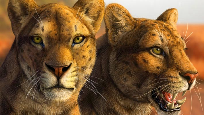
Le premier chat à dents de sabre obtient un nouveau visage après des décennies de mystère
dent paléoart reconstitution États-Unis Pliocène mammifères crâne
Un crâne presque complet découvert il y a plusieurs décennies en Arizona a donné aux paléontologues leur aperçu le plus clair à ce jour d'Adelphailurus kansensis, une espèce énigmatique de félidés qui habitait l'Amérique du Nord il y a plus de 5 millions d'années et occupait une des premières branches de l'arbre généalogique des chats à dents de sabre. L'article Le premier chat à dents de sabre obtient un nouveau visage après des décennies de mystère est apparu en premier sur Sci.News : Breaking Science News.
29/06/2026 sci-news ⚙ Traduction automatique
Que mangeaient les dinosaures ? Un guide utile sur les régimes alimentaires des dinosaures
os mâchoire dent fossile Dinosauria étude
Vous êtes-vous déjà demandé ce que mangeaient les dinosaures ? C’est l’une des questions les plus fréquemment posées chez Everything Dinosaur. La réponse est étonnamment variée. Certains dinosaures étaient de féroces mangeurs de viande, tandis que d’autres se nourrissaient de plantes. D'autres étaient omnivores (mangant à la fois des plantes et de la viande). Les scientifiques étudient les dents fossiles, les os de la mâchoire, le contenu de l'estomac et même les fossiles
22/06/2026 everythingdinosaur ⚙ Traduction automatique
1 2 3 4 5 6 7 8