←

crâne

Type de vue

215 image(s) · 30 Actualités

Galerie d'images

Nemicolopterus crypticus holotype (IVPP V-14377).

(A) Skeleton overview, and (B) schematic drawing. (C) Skull (right lateral view), and (D) schematic drawing. Abbreviations: cdv, caudal vertebrae; co, coracoid; cv, cervical vertebra; d1–d4, digits 1–4; f, frontal; fe, femur; h, humerus; hy, hyoid; il, illium; is, ischium; j, jugal; l, left; la, lacrimal; mand, mandible; mc, metacarpal; mt, metatarsal; naof, nasoantorbital fenestra; or, orbit; pa, parietal; pm, premaxilla; ph, phalanx; ti, tibia; r, right; sca, scapula. Scale bars: A–B, 100 mm; C–D, 5 mm.
Taxons Nemicolopterus

Nemicolopterus crypticus holotype (IVPP V-14377). (A) Skeleton overview, and (B) schematic drawing. (C) Skull (right lateral view), and (D) schematic drawing. Abbreviations: cdv, caudal vertebrae; co, coracoid; cv, cervical vertebra; d1–d4, digits 1–4; f, frontal; fe, femur; h, humerus; hy, hyoid; il, illium; is, ischium; j, jugal; l, left; la, lacrimal; mand, mandible; mc, metacarpal; mt, metatarsal; naof, nasoantorbital fenestra; or, orbit; pa, parietal; pm, premaxilla; ph, phalanx; ti, tibia; r, right; sca, scapula. Scale bars: A–B, 100 mm; C–D, 5 mm.

humérus écaille vertèbre dessin +4
Udelartitan is a titanosaurian sauropod from the Late Cretaceous of what is now Uruguay. It had the typical sauropod body plan, standing on four thick pillar-like legs, and possessing a long neck. Titanosaurs are a very diverse group, Udelartitan was a small one, estimated at only 15 m in body length. Though a skull has yet to be discovered, it probably had peglike teeth, and a large gut for processing vegetation.
Taxons Udelartitan

Udelartitan is a titanosaurian sauropod from the Late Cretaceous of what is now Uruguay. It had the typical sauropod body plan, standing on four thick pillar-like legs, and possessing a long neck. Titanosaurs are a very diverse group, Udelartitan was a small one, estimated at only 15 m in body length. Though a skull has yet to be discovered, it probably had peglike teeth, and a large gut for processing vegetation.

Uruguay Crétacé Crétacé supérieur Titanosauria +2
Ludodactylus sibbicki. A, B, holotype (SMNK PAL 3828) skull and lower jaw in right lateral view.
Taxons Ludodactylus

Ludodactylus sibbicki. A, B, holotype (SMNK PAL 3828) skull and lower jaw in right lateral view.

holotype Ludodactylus crâne
Reconstruction of the skull of Spinops sternbergorum gen. et sp. nov. from the Campanian of Dinosaur Provincial Park, southern Alberta, in right lateral view. Preserved elements are stippled; missing portions are dotted and modeled after Centrosaurus apertus.
Taxons Spinops

Reconstruction of the skull of Spinops sternbergorum gen. et sp. nov. from the Campanian of Dinosaur Provincial Park, southern Alberta, in right lateral view. Preserved elements are stippled; missing portions are dotted and modeled after Centrosaurus apertus.

Campanien Centrosaurus Dinosauria Spinops +1
Skull of troodontid theropod Xixiasaurus henanensis gen. et sp. nov. (HGM 41HIII−0201; holotype), lower–middle Majiacun Formation (Upper Cretaceous: Coniacian–Campanian), Henan Province, China; in dorsal (A); lateral (B), and ventral (C) views.

Note that this version of the image does not include the interpretative lines of the version in the paper.
Taxons Xixiasaurus

Skull of troodontid theropod Xixiasaurus henanensis gen. et sp. nov. (HGM 41HIII−0201; holotype), lower–middle Majiacun Formation (Upper Cretaceous: Coniacian–Campanian), Henan Province, China; in dorsal (A); lateral (B), and ventral (C) views. Note that this version of the image does not include the interpretative lines of the version in the paper.

Chine Campanien Coniacien Crétacé +5
Skull diagram featuring the holotype skull (ROM 1790) of the ornithomimid Rativates evadens, from the Late Cretaceous period. Abbreviations: d, dentary; f, frontal; l, lacrimal; m, maxilla; n, nasal; pm, premaxila; ps, paraphenoid.
Taxons Rativates

Skull diagram featuring the holotype skull (ROM 1790) of the ornithomimid Rativates evadens, from the Late Cretaceous period. Abbreviations: d, dentary; f, frontal; l, lacrimal; m, maxilla; n, nasal; pm, premaxila; ps, paraphenoid.

Crétacé Crétacé supérieur holotype Ornithomimidae +2
Reconstructed skull of Archaeornithoides, known material in brown.
Taxons Archaeornithoides

Reconstructed skull of Archaeornithoides, known material in brown.

Archaeornithoides crâne
Holotype skull of Wannanosaurus yansiensis on display at the Paleozoological Museum of China.
Taxons Wannanosaurus

Holotype skull of Wannanosaurus yansiensis on display at the Paleozoological Museum of China.

musée Chine holotype Wannanosaurus +1
Holotype specimen (PIMUZ A/III 1274) of Prosantosaurus scheffoldi gen. et spec. nov. from the upper Prosanto Formation (Early Ladinian, Middle Triassic) of Ducanfurgga locality no. 4, southwest of Davos, Canton of Grisons, south-eastern Switzerland. A Nearly complete specimen as prepared in dorsal view. The posterior part of the tail was lost prior to burial. Both forearms are not visible but lie below the trunk region, pointing in an anteromedial direction (see Additional file 1: Fig. S3A). B Detail of skull and anterior neck region. C Outline sketch of skull sutures. D Detail of shoulder girdle (claviculae, scapulae) and anterior dorsal vertebrae and ribs. E Detail of right humerus. F Detail of posterior dorsal vertebrae and ribs, sacral vertebrae and ribs, and anterior caudal vertebrae and ribs. G Detail of left ilium and hindlimb. ar articular; as astragalus; bo basioccipital; cal calcaneus; cl clavicula; co coracoid; d dentary; en external naris; eo exoccipital; fe femur; fi fibula; fr frontal; hu humerus; il ilium; in internal naris; is ischium; j jugal; mx maxilla; na naris; o orbit; pa parietal; pl palatine; pmx premaxilla; pof postfrontal; po postorbital; prf prefrontal; pt pterygoid; pu pubis; q quadrate; qj quadratojugal; ti tibia; sacr sacral rib; sc scapula; so supraoccipital; sp splenial; sq squamosal; su surangular; utf upper temporal fenestra; v vomer
Taxons Prosantosaurus

Holotype specimen (PIMUZ A/III 1274) of Prosantosaurus scheffoldi gen. et spec. nov. from the upper Prosanto Formation (Early Ladinian, Middle Triassic) of Ducanfurgga locality no. 4, southwest of Davos, Canton of Grisons, south-eastern Switzerland. A Nearly complete specimen as prepared in dorsal view. The posterior part of the tail was lost prior to burial. Both forearms are not visible but lie below the trunk region, pointing in an anteromedial direction (see Additional file 1: Fig. S3A). B Detail of skull and anterior neck region. C Outline sketch of skull sutures. D Detail of shoulder girdle (claviculae, scapulae) and anterior dorsal vertebrae and ribs. E Detail of right humerus. F Detail of posterior dorsal vertebrae and ribs, sacral vertebrae and ribs, and anterior caudal vertebrae and ribs. G Detail of left ilium and hindlimb. ar articular; as astragalus; bo basioccipital; cal calcaneus; cl clavicula; co coracoid; d dentary; en external naris; eo exoccipital; fe femur; fi fibula; fr frontal; hu humerus; il ilium; in internal naris; is ischium; j jugal; mx maxilla; na naris; o orbit; pa parietal; pl palatine; pmx premaxilla; pof postfrontal; po postorbital; prf prefrontal; pt pterygoid; pu pubis; q quadrate; qj quadratojugal; ti tibia; sacr sacral rib; sc scapula; so supraoccipital; sp splenial; sq squamosal; su surangular; utf upper temporal fenestra; v vomer

humérus Suisse Ladinien Trias moyen +6
Reconstructed skull of the turiasaurian Mierasaurus, based on the holotype UMNH.VP.26004.
Taxons Mierasaurus

Reconstructed skull of the turiasaurian Mierasaurus, based on the holotype UMNH.VP.26004.

holotype Mierasaurus Turiasauria crâne
Riparovenator milnerae holotype skull
Taxons Riparovenator

Riparovenator milnerae holotype skull

holotype Riparovenator crâne
Caihong juji holotype specimen (PMoL-B00175). Photographs of the slab (a) and counter slab (b) and line drawing (c) of the specimen based on both slabs. Photograph (d) and line drawing (e) of a composite of the rostrum of the skull and mandible exposed on the counter slab and the post-rostrum cranium exposed on the slab. Arrows indicate lacrimal crests. Question mark indicates uncertain identification. Scale bars: 10 cm a–c, 1 cm d and e. aof antorbital fenestra, cav caudal vertebra, cev cervical vertebra, dr dorsal rib, dv dorsal vertebra, ect ectopterygoid, emf external mandibular fenestra, en external naris, f feather, fu furcula, ga gastralia, hy hyoid, il ilium, is ischium, la left angular, lar left articular, lc left coracoid, lcr lacrimal crest, ld left dentary, lf left, frontal, lfe left femur, lh left humerus, lj left jugal, ll left lacrimal, lma left maxilla, lm left manus, ln left nasal, lp left pes, lpa left palatine, lpo left postorbital, lq left quadrate, lqj left quadratojugal, lr left radius, ls left scapula, lsp left splenial, lsa left surangular, lsq left squamosal, lt left tibiotarsus, lu left ulna, ma mandible, mf maxillary fenestra, o orbit, p parietal, pm premaxilla, pt pterygoid, pu pubis, rar right articular, rc right coracoid, rd right dentary, rfe right femur, rh right humerus, rm right manus, rp right pes, rpra right prearticular, rq right quadrate, rr right radius, rs right scapula, rt right tibiotarsus, ru right ulna, scl sclerotic bones, sk skull, sy synsacrum
Taxons Caihong

Caihong juji holotype specimen (PMoL-B00175). Photographs of the slab (a) and counter slab (b) and line drawing (c) of the specimen based on both slabs. Photograph (d) and line drawing (e) of a composite of the rostrum of the skull and mandible exposed on the counter slab and the post-rostrum cranium exposed on the slab. Arrows indicate lacrimal crests. Question mark indicates uncertain identification. Scale bars: 10 cm a–c, 1 cm d and e. aof antorbital fenestra, cav caudal vertebra, cev cervical vertebra, dr dorsal rib, dv dorsal vertebra, ect ectopterygoid, emf external mandibular fenestra, en external naris, f feather, fu furcula, ga gastralia, hy hyoid, il ilium, is ischium, la left angular, lar left articular, lc left coracoid, lcr lacrimal crest, ld left dentary, lf left, frontal, lfe left femur, lh left humerus, lj left jugal, ll left lacrimal, lma left maxilla, lm left manus, ln left nasal, lp left pes, lpa left palatine, lpo left postorbital, lq left quadrate, lqj left quadratojugal, lr left radius, ls left scapula, lsp left splenial, lsa left surangular, lsq left squamosal, lt left tibiotarsus, lu left ulna, ma mandible, mf maxillary fenestra, o orbit, p parietal, pm premaxilla, pt pterygoid, pu pubis, rar right articular, rc right coracoid, rd right dentary, rfe right femur, rh right humerus, rm right manus, rp right pes, rpra right prearticular, rq right quadrate, rr right radius, rs right scapula, rt right tibiotarsus, ru right ulna, scl sclerotic bones, sk skull, sy synsacrum

os crête plume humérus +8
Figure 4: Anterior portion of the skull of Dakotadon lakotaensis (SDSM 8656).
(A) photograph in left lateral view; (B) photograph in right lateral view; (C) photograph in ventral view. Abbreviations: ant, anterior; post, posterior; vent, ventral. Scale bars equal 5.0 cm.
Taxons Dakotadon

Figure 4: Anterior portion of the skull of Dakotadon lakotaensis (SDSM 8656). (A) photograph in left lateral view; (B) photograph in right lateral view; (C) photograph in ventral view. Abbreviations: ant, anterior; post, posterior; vent, ventral. Scale bars equal 5.0 cm.

écaille Dakotadon crâne
Ceratosuchops inferodios Holotype skull fragments
Taxons Ceratosuchops

Ceratosuchops inferodios Holotype skull fragments

holotype Ceratosuchops crâne
Partial skull and anterior cervical vertebrae of unidentified pliosaurid plesiosaurian (MPPL 18797) from the uppermost Callovian–middle Oxfordian of Kaberlaba (Asiago, Italy), in left lateral view. Photograph (A) and explanatory drawing (B). White, preserved bone surfaces; dark grey, preserved margins of skull fenestrae; light grey, eroded bone surface; cross hatching, broken bone.
Taxons Anguanax

Partial skull and anterior cervical vertebrae of unidentified pliosaurid plesiosaurian (MPPL 18797) from the uppermost Callovian–middle Oxfordian of Kaberlaba (Asiago, Italy), in left lateral view. Photograph (A) and explanatory drawing (B). White, preserved bone surfaces; dark grey, preserved margins of skull fenestrae; light grey, eroded bone surface; cross hatching, broken bone.

os dessin Italie Callovien +6
Identifier: annalsofmedicalh01pack (find matches)
Title: Annals of medical history
Year: 1917 (1910s)
Authors:  Packard, Francis R. (Francis Randolph), 1870-1950
Subjects:  Medicine
Publisher:  New York P.B. Hoeber
Contributing Library:  Gerstein - University of Toronto
Digitizing Sponsor:  University of Toronto

View Book Page: Book Viewer
About This Book: Catalog Entry
View All Images: All Images From Book
Click here to view book online to see this illustration in context in a browseable online version of this book.

Text Appearing Before Image:
ail strongly Hexed, and the toescontracted and appressed. The whole atti-tude strongly suggests a spastic distress,possibly brought on by some form ofpoisoning of the central nervous system,from infection or the deglutition of somepoisonous substance. 14. Osteomalacia is evidently the causeof the hypertrophy of the bones of Lim-nocyron potens, an early carnivore from theWashakie Eocene of Wyoming, nearly3,000,000 years old. MATERIALS AND METHODS The material described in the presentpaper has been loaned the writer lor de-scription by the Field Museum of Chii Osborn: Hull. Amcr. \lus. Natl. Hist., 1917,vol. 35. !>• 733. P>- 28. Studies in Paleopathology 393 by the American Museum of Natural His-tory of New York City, by Walker Museumof the University of Chicago, and by the Uni-versity of Kansas Natural History Museum. made by the well-known petrographic meth-ods so common in all geological labora-tories. The diagnoses, where they are at-tempted, arc made from comparisons of the
Text Appearing After Image:
Fig. 20. The skeleton of Strutbiomimus altus, a small dinosaur from the BeliyRiver series (Cretaceous), Red Deer River, Alberta, Canada, now regarded as ofapproximately the same age as the Judith River series. The unique feature ofthe skull is the total absence of teeth, with a size of skull one-third larger thanthe ostrich and a length of body of about fifteen feet. The position of the skeletonis decidedly that of the opisthotonos which may be regarded as an indication ofdisease. (After Osborn). A beautiful specimen of an osteoma, theonly one known so far, on the vertebra of aKansas Cretaceous mosasaur, was given thewriter by Dr. J. M. Armstrong of St. Paul.The writer expresses his obligations to thegentlemen connected with the above-men-tioned institutions and to Dr. Armstrong.The methods used are a combination ofprocedures in the various lines involved.Microscopic sections, which can be madethin enough for immersion lens study, are material with similar lesions in recent hu-man mat

Note About Images

Please note that these images are extracted from scanned page images that may have been digitally enhanced for readability - coloration and appearance of these illustrations may not perfectly resemble the original work.
Taxons Hexing

Identifier: annalsofmedicalh01pack (find matches) Title: Annals of medical history Year: 1917 (1910s) Authors: Packard, Francis R. (Francis Randolph), 1870-1950 Subjects: Medicine Publisher: New York P.B. Hoeber Contributing Library: Gerstein - University of Toronto Digitizing Sponsor: University of Toronto View Book Page: Book Viewer About This Book: Catalog Entry View All Images: All Images From Book Click here to view book online to see this illustration in context in a browseable online version of this book. Text Appearing Before Image: ail strongly Hexed, and the toescontracted and appressed. The whole atti-tude strongly suggests a spastic distress,possibly brought on by some form ofpoisoning of the central nervous system,from infection or the deglutition of somepoisonous substance. 14. Osteomalacia is evidently the causeof the hypertrophy of the bones of Lim-nocyron potens, an early carnivore from theWashakie Eocene of Wyoming, nearly3,000,000 years old. MATERIALS AND METHODS The material described in the presentpaper has been loaned the writer lor de-scription by the Field Museum of Chii Osborn: Hull. Amcr. \lus. Natl. Hist., 1917,vol. 35. !>• 733. P>- 28. Studies in Paleopathology 393 by the American Museum of Natural His-tory of New York City, by Walker Museumof the University of Chicago, and by the Uni-versity of Kansas Natural History Museum. made by the well-known petrographic meth-ods so common in all geological labora-tories. The diagnoses, where they are at-tempted, arc made from comparisons of the Text Appearing After Image: Fig. 20. The skeleton of Strutbiomimus altus, a small dinosaur from the BeliyRiver series (Cretaceous), Red Deer River, Alberta, Canada, now regarded as ofapproximately the same age as the Judith River series. The unique feature ofthe skull is the total absence of teeth, with a size of skull one-third larger thanthe ostrich and a length of body of about fifteen feet. The position of the skeletonis decidedly that of the opisthotonos which may be regarded as an indication ofdisease. (After Osborn). A beautiful specimen of an osteoma, theonly one known so far, on the vertebra of aKansas Cretaceous mosasaur, was given thewriter by Dr. J. M. Armstrong of St. Paul.The writer expresses his obligations to thegentlemen connected with the above-men-tioned institutions and to Dr. Armstrong.The methods used are a combination ofprocedures in the various lines involved.Microscopic sections, which can be madethin enough for immersion lens study, are material with similar lesions in recent hu-man mat Note About Images Please note that these images are extracted from scanned page images that may have been digitally enhanced for readability - coloration and appearance of these illustrations may not perfectly resemble the original work.

os vertèbre livre musée +11
1 2 3 4 5 6 7 8 9 10 11 12 13 14

Actualités

Un nouveau genre de dinosaure troodontidé du Nouveau-Mexique
os Mexique Campanien Crétacé Crétacé supérieur fossile Dinevenator Dinosauria Troodontidae nouvelle espèce crâne
Les paléontologues ont nommé une nouvelle espèce de dinosaure théropode du Nouveau-Mexique. Le dinosaure, Dinevenator Robustus, vivait il y a environ 73 millions d'années (stade faunique campanien du Crétacé supérieur). Les chercheurs ont identifié le dinosaure à partir d’un os fossile du crâne (un frontal) trouvé dans le nord-ouest du Nouveau-Mexique. Il représente un nouveau genre et une nouvelle espèce de troodontidé. Troodontidés
13/08/2026 everythingdinosaur ⚙ Traduction automatique
L’oiseau dodo possédait un monde sensoriel secret que les scientifiques découvrent seulement maintenant
oiseau tomographie découverte crâne
Les tomodensitogrammes des crânes de dodo les plus rares au monde suggèrent que l'oiseau disparu aurait pu avoir un odorat plus fort, un bec très sensible et un mode de vie qui s'étendait jusqu'à l'aube et au crépuscule. Ces découvertes contribuent à renverser des mythes vieux de plusieurs siècles et à révéler un animal bien plus complexe que le fameux dodo « stupide » de l’imagination populaire.
09/08/2026 sciencedaily-paleo ⚙ Traduction automatique
Un fossile oublié vient de révéler un nouveau prédateur du Trias datant d'il y a 210 millions d'années
mâchoire prédateur proie Mexique Trias fossile crâne
Un fossile négligé depuis près de 80 ans a révélé un parent de crocodile jusqu'alors inconnu qui vivait il y a 210 millions d'années dans ce qui est aujourd'hui le Nouveau-Mexique. Nommé Eosphorosuchus lacrimosa, ce prédateur de la taille d'un chacal avait un crâne renforcé, des muscles de mâchoire puissants et un museau court conçu pour s'attaquer à des proies plus grosses.
30/07/2026 sciencedaily ⚙ Traduction automatique
Ce fossile de dinosaure capture les derniers instants d'une attaque de T. rex
dent prédateur proie fossile Dinosauria Edmontosaurus Tyrannosaurus découverte crâne
Un crâne fossilisé d'Edmontosaure avec une dent de tyrannosaure encore incrustée dans son visage a fourni aux scientifiques de rares preuves d'une rencontre dramatique entre prédateur et proie. La découverte suggère que le carnivore géant a prononcé une morsure face-à-face incroyablement puissante, offrant de nouveaux indices sur la façon dont le tyrannosaure chassait.
14/07/2026 sciencedaily ⚙ Traduction automatique
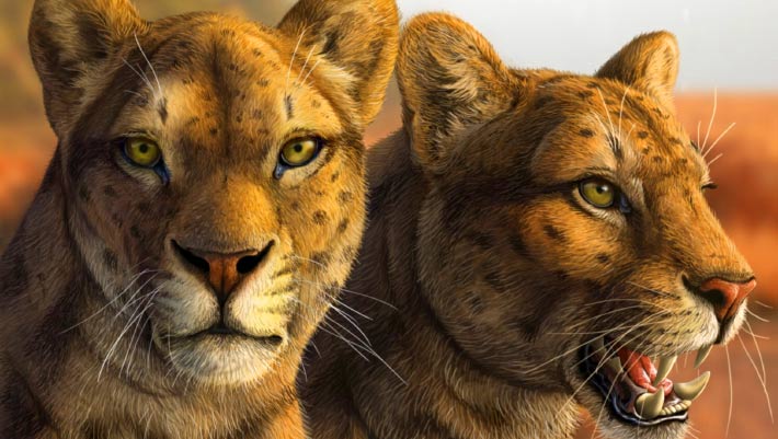
Early Saber-Toothed Cat Gets New Face after Decades of Mystery
Le premier chat à dents de sabre obtient un nouveau visage après des décennies de mystère
dent paléoart reconstitution États-Unis Pliocène mammifères crâne
Un crâne presque complet découvert il y a plusieurs décennies en Arizona a donné aux paléontologues leur aperçu le plus clair à ce jour d'Adelphailurus kansensis, une espèce énigmatique de félidés qui habitait l'Amérique du Nord il y a plus de 5 millions d'années et occupait une des premières branches de l'arbre généalogique des chats à dents de sabre. L'article Le premier chat à dents de sabre obtient un nouveau visage après des décennies de mystère est apparu en premier sur Sci.News : Breaking Science News.
29/06/2026 sci-news ⚙ Traduction automatique
1 2 3 4 5 6