crâne

Type de vue

244 image(s) · 27 Actualités

Galerie d'images

The skull of Zupaysaurus rougieri (A) in right lateral view. Scale bar equals 5 cm. Cropped from Figure 2 in the source.
anfe - antorbital fenestra
emf - external mandibular fenestra
itf - infratemporal fenestra
or - orbit
Taxons Zupaysaurus

The skull of Zupaysaurus rougieri (A) in right lateral view. Scale bar equals 5 cm. Cropped from Figure 2 in the source. anfe - antorbital fenestra emf - external mandibular fenestra itf - infratemporal fenestra or - orbit

écaille Zupaysaurus crâne
Skull diagram featuring the holotype skull (ROM 1790) of the ornithomimid Rativates evadens, from the Late Cretaceous period. Abbreviations: d, dentary; f, frontal; l, lacrimal; m, maxilla; n, nasal; pm, premaxila; ps, paraphenoid.
Taxons Rativates

Skull diagram featuring the holotype skull (ROM 1790) of the ornithomimid Rativates evadens, from the Late Cretaceous period. Abbreviations: d, dentary; f, frontal; l, lacrimal; m, maxilla; n, nasal; pm, premaxila; ps, paraphenoid.

Crétacé Crétacé supérieur holotype Ornithomimidae +2
Mounted skull of EMK 0012 (Lokiceratops rangiformis holotype). (A) Mounted skull in posterior view. (B) Mounted skull in right lateral view. (C) Mounted skull in dorsal view. (D) Mounted skull in left lateral view. Areas in gray are reconstructed. Minor changes from side to side and in the orbits are the result of post-depositional deformation. Photos contain parallax. Scale bar equals 1 m.
Taxons Lokiceratops

Mounted skull of EMK 0012 (Lokiceratops rangiformis holotype). (A) Mounted skull in posterior view. (B) Mounted skull in right lateral view. (C) Mounted skull in dorsal view. (D) Mounted skull in left lateral view. Areas in gray are reconstructed. Minor changes from side to side and in the orbits are the result of post-depositional deformation. Photos contain parallax. Scale bar equals 1 m.

écaille holotype Lokiceratops crâne
Reconstruction of the skull of Spinops sternbergorum gen. et sp. nov. from the Campanian of Dinosaur Provincial Park, southern Alberta, in right lateral view. Preserved elements are stippled; missing portions are dotted and modeled after Centrosaurus apertus.
Taxons Spinops

Reconstruction of the skull of Spinops sternbergorum gen. et sp. nov. from the Campanian of Dinosaur Provincial Park, southern Alberta, in right lateral view. Preserved elements are stippled; missing portions are dotted and modeled after Centrosaurus apertus.

Campanien Centrosaurus Dinosauria Spinops +1
Photograph of the holotype skull of Titanoceratops ouranos, from the late Campanian aged Kirtland/Fruitland beds of New Mexico, USA. Photograph by Nick Longrich
Taxons Titanoceratops

Photograph of the holotype skull of Titanoceratops ouranos, from the late Campanian aged Kirtland/Fruitland beds of New Mexico, USA. Photograph by Nick Longrich

Mexique États-Unis Fruitland Kirtland +4
Ceratopsipes goldenensis Lockley & Hunt, 1995 - ceratopsian dinosaur trackway in the Cretaceous of Colorado, USA.
Ceratopsians are the "horned dinosaurs".  They were large, quadrupedal, herbivorous dinosaurs having a beaked skull and a frill - an extension of bone behind the skull that partially covered the neck.  Ceratopsian dinosaurs are known from the Jurassic and Cretaceous.  The last members of the group died out at the Cretaceous-Tertiary boundary, 65 million years ago.
Seen here is a ceratopsian dinosaur trackway.  Ceratopsian footprints are very rare.  The best examples are in Colorado's Laramie Formation, a nonmarine, coastal plain to deltaic succession of mostly mixed siliciclastic sedimentary rocks - sandstones, siltstones, claystones, and coals.  The footprints here consist of convex bulges on the basal surfaces of structurally-tilted sandstone beds.
The ichnospecies Ceratopsipes goldenensis was named based on footprints at this locality.  The trackmaker was very likely Triceratops, the # 1 most famous ceratopsian dinosaur.


From on-site signage:
Triceratops Tracks
Several tracks of Triceratops, or a closely related horned dinosaur were first discovered in this area and named Ceratopsipes goldenensis (meaning tracks of a horned dinosaur from Golden).  By happy coincidence, the first Triceratops known to science also comes from the greater Denver area near 13th Avenue and Federal Blvd.  Growing to a length of almost 25 feet, Triceratops and other members of the horned dinosaur family were herbivorous animals that roamed in herds.  Their trademark horns and frills that covered their necks probably served for defense.  A replica Triceratops skull is on display in the clubhouse at the Fossil Trace Golf Club.
These tracks (identified by small signs) help us understand that Triceratops walked with a narrow gait and erect front limbs  than with a wide, sprawling gait as previously depicted.  In 1887, the first Triceratops bones known to science were also discovered locally at a site near 13th Avenue and Federal Boulevard in Denver.
Nearby one may see many other traces of life such as burrows, probably made by invertebrates and impressions of plant debris.  Note that you are looking at all tracks and traces from the underside - in negative aspect - what one might call a worm’s eye view.  All of these trace fossils indicate that the Golden area once had a warm, temperate to subtropical climate.


Classification: Animalia, Chordata, Vertebrata, Reptilia, Archosauria, Dinosauria, Ornithischia, Marginocephalia, Ceratopsia, Ceratopsidae
Stratigraphy: Laramie Formation, Maastrichtian Stage, upper Upper Cretaceous
Locality: outcrop along Triceratops Trail, Parfet Prehistoric Preserve, southern side of the town of Golden, Colorado, USA (~vicinity of 39° 44' 35.24" North latitude, 105° 13’ 09.69" West longitude)


Some info. from:

Lockley & Hunt (1995) - Ceratopsid tracks and associated ichnofauna from the Laramie Formation (Upper Cretaceous: Maastrichtian) of Colorado.  Journal of Vertebrate Paleontology 15: 592-614.
Taxons Ceratopsipes

Ceratopsipes goldenensis Lockley & Hunt, 1995 - ceratopsian dinosaur trackway in the Cretaceous of Colorado, USA. Ceratopsians are the "horned dinosaurs". They were large, quadrupedal, herbivorous dinosaurs having a beaked skull and a frill - an extension of bone behind the skull that partially covered the neck. Ceratopsian dinosaurs are known from the Jurassic and Cretaceous. The last members of the group died out at the Cretaceous-Tertiary boundary, 65 million years ago. Seen here is a ceratopsian dinosaur trackway. Ceratopsian footprints are very rare. The best examples are in Colorado's Laramie Formation, a nonmarine, coastal plain to deltaic succession of mostly mixed siliciclastic sedimentary rocks - sandstones, siltstones, claystones, and coals. The footprints here consist of convex bulges on the basal surfaces of structurally-tilted sandstone beds. The ichnospecies Ceratopsipes goldenensis was named based on footprints at this locality. The trackmaker was very likely Triceratops, the # 1 most famous ceratopsian dinosaur. From on-site signage: Triceratops Tracks Several tracks of Triceratops, or a closely related horned dinosaur were first discovered in this area and named Ceratopsipes goldenensis (meaning tracks of a horned dinosaur from Golden). By happy coincidence, the first Triceratops known to science also comes from the greater Denver area near 13th Avenue and Federal Blvd. Growing to a length of almost 25 feet, Triceratops and other members of the horned dinosaur family were herbivorous animals that roamed in herds. Their trademark horns and frills that covered their necks probably served for defense. A replica Triceratops skull is on display in the clubhouse at the Fossil Trace Golf Club. These tracks (identified by small signs) help us understand that Triceratops walked with a narrow gait and erect front limbs than with a wide, sprawling gait as previously depicted. In 1887, the first Triceratops bones known to science were also discovered locally at a site near 13th Avenue and Federal Boulevard in Denver. Nearby one may see many other traces of life such as burrows, probably made by invertebrates and impressions of plant debris. Note that you are looking at all tracks and traces from the underside - in negative aspect - what one might call a worm’s eye view. All of these trace fossils indicate that the Golden area once had a warm, temperate to subtropical climate. Classification: Animalia, Chordata, Vertebrata, Reptilia, Archosauria, Dinosauria, Ornithischia, Marginocephalia, Ceratopsia, Ceratopsidae Stratigraphy: Laramie Formation, Maastrichtian Stage, upper Upper Cretaceous Locality: outcrop along Triceratops Trail, Parfet Prehistoric Preserve, southern side of the town of Golden, Colorado, USA (~vicinity of 39° 44' 35.24" North latitude, 105° 13’ 09.69" West longitude) Some info. from: Lockley & Hunt (1995) - Ceratopsid tracks and associated ichnofauna from the Laramie Formation (Upper Cretaceous: Maastrichtian) of Colorado. Journal of Vertebrate Paleontology 15: 592-614.

os États-Unis Crétacé Jurassique +6
Hualianceratops holotype IVVP V18641, Skull reconstruction
Taxons Hualianceratops

Hualianceratops holotype IVVP V18641, Skull reconstruction

holotype Hualianceratops crâne
Figure 1: Holotype and skeletal reconstruction of Mosaiceratops azumai, gen. et sp. nov (ZMNH M8856). (a) photograph and line drawing of ZMNH M8856; (b) skeletal reconstruction showing preserved elements in white. Scale bar 10 cm. Abbreviations: a, astragalus; boc, basioccipital; c, calcaneum; cav, caudal vertebra; ch, chevron; cv, cervical vertebra; dr, dorsal rib; dv, dorsal vertebra; f, frontal; fem, femur; fl, fibula; h, humerus; il, ilium; is, ischium; L, left; mt, metatarsal; ph, phalanx/phalanges; po, postorbital; R, right; sk, skull; sq, squamosal; t, tibia; td, tendon; ?, undiagnostic remains.
Taxons Mosaiceratops

Figure 1: Holotype and skeletal reconstruction of Mosaiceratops azumai, gen. et sp. nov (ZMNH M8856). (a) photograph and line drawing of ZMNH M8856; (b) skeletal reconstruction showing preserved elements in white. Scale bar 10 cm. Abbreviations: a, astragalus; boc, basioccipital; c, calcaneum; cav, caudal vertebra; ch, chevron; cv, cervical vertebra; dr, dorsal rib; dv, dorsal vertebra; f, frontal; fem, femur; fl, fibula; h, humerus; il, ilium; is, ischium; L, left; mt, metatarsal; ph, phalanx/phalanges; po, postorbital; R, right; sk, skull; sq, squamosal; t, tibia; td, tendon; ?, undiagnostic remains.

humérus écaille vertèbre dessin +3
Skeletal diagram featuring the optimal remains of the holotype of Graciliceratops mongoliensis: ZPAL MgD-I/156.[1] Found in the Bayan Shireh Formation, in the original description the remains were referred to the genus Microceratops (now obsolete).[2] However, Sereno in 2000 noted that there was no base for this referral, then, he created a new genus and species for this specimen.[1] The holotype is very fragmented (specially the skull), consisting of:[2]
Fragmented skull; 4 cervical, 12 dorsal and 7 sacral vertebrae; right scapula; proximal end of left scapula; left coracoid; right humerus, radius and fragmentary ulna; proximal and distal end of left humerus; proximal fragments of both pubis; fragments of both illium and fragment of right ischium; right femur, tibia and nearly complete pes; distal part of left tibia, fragmentary left pes; tarsals and isolated ribs.[2]

The sacral vertebrae are not fused, an indicator of the immaturity of this specimen; the estimated adult size is about 2 meters long or similar to Protoceratops.[2][1] Right quadratojugal, quadrate and fragmentary jugal were reversed in order to get an optimal view.
Taxons Graciliceratops

Skeletal diagram featuring the optimal remains of the holotype of Graciliceratops mongoliensis: ZPAL MgD-I/156.[1] Found in the Bayan Shireh Formation, in the original description the remains were referred to the genus Microceratops (now obsolete).[2] However, Sereno in 2000 noted that there was no base for this referral, then, he created a new genus and species for this specimen.[1] The holotype is very fragmented (specially the skull), consisting of:[2] Fragmented skull; 4 cervical, 12 dorsal and 7 sacral vertebrae; right scapula; proximal end of left scapula; left coracoid; right humerus, radius and fragmentary ulna; proximal and distal end of left humerus; proximal fragments of both pubis; fragments of both illium and fragment of right ischium; right femur, tibia and nearly complete pes; distal part of left tibia, fragmentary left pes; tarsals and isolated ribs.[2] The sacral vertebrae are not fused, an indicator of the immaturity of this specimen; the estimated adult size is about 2 meters long or similar to Protoceratops.[2][1] Right quadratojugal, quadrate and fragmentary jugal were reversed in order to get an optimal view.

humérus description holotype spécimen +4
Holotype skull of Wannanosaurus yansiensis on display at the Paleozoological Museum of China.
Taxons Wannanosaurus

Holotype skull of Wannanosaurus yansiensis on display at the Paleozoological Museum of China.

musée Chine holotype Wannanosaurus +1
Diagram featuring the holotype skull (ZPAL MgD-I/105) of Tylocephale gilmorei in left lateral view. Scale bar equals 5 cm (50 mm). Based on Maryańska & Osmólska, 1974,[1] and Sullivan, 2006.[2]
References

↑ (1974). "Pachycephalosauria, a new suborder of ornithischian dinosaurs". Palaeontologica Polonica (30): 45−102.

↑ (2006). "A taxonomic review of the Pachycephalosauridae (Dinosauria: Ornithischia)". New Mexico Museum of Natural History and Science Bulletin (35): 347–365.
Taxons Tylocephale

Diagram featuring the holotype skull (ZPAL MgD-I/105) of Tylocephale gilmorei in left lateral view. Scale bar equals 5 cm (50 mm). Based on Maryańska & Osmólska, 1974,[1] and Sullivan, 2006.[2] References ↑ (1974). "Pachycephalosauria, a new suborder of ornithischian dinosaurs". Palaeontologica Polonica (30): 45−102. ↑ (2006). "A taxonomic review of the Pachycephalosauridae (Dinosauria: Ornithischia)". New Mexico Museum of Natural History and Science Bulletin (35): 347–365.

écaille musée Mexique holotype +6
Lateral diagram of the skull of Sinocephale bexelli (Bohlin, 1953) with unknown regions (grey) restored after Stegoceras
Taxons Sinocephale

Lateral diagram of the skull of Sinocephale bexelli (Bohlin, 1953) with unknown regions (grey) restored after Stegoceras

Sinocephale Stegoceras crâne
Holotype of Transylvanosaurus platycephalus, LPB (FGGUB) R.2070, a partial skull comprising the articulated basicranium and both frontals. Scale bar: 1 cm.
Taxons Transylvanosaurus

Holotype of Transylvanosaurus platycephalus, LPB (FGGUB) R.2070, a partial skull comprising the articulated basicranium and both frontals. Scale bar: 1 cm.

écaille holotype Transylvanosaurus partiel +1
skull of iguanodontian Altirhinus kurzanovi
Taxons Altirhinus

skull of iguanodontian Altirhinus kurzanovi

Altirhinus Iguanodontia crâne
Figure 4: Anterior portion of the skull of Dakotadon lakotaensis (SDSM 8656).
(A) photograph in left lateral view; (B) photograph in right lateral view; (C) photograph in ventral view. Abbreviations: ant, anterior; post, posterior; vent, ventral. Scale bars equal 5.0 cm.
Taxons Dakotadon

Figure 4: Anterior portion of the skull of Dakotadon lakotaensis (SDSM 8656). (A) photograph in left lateral view; (B) photograph in right lateral view; (C) photograph in ventral view. Abbreviations: ant, anterior; post, posterior; vent, ventral. Scale bars equal 5.0 cm.

écaille Dakotadon crâne
Angulomastacator skull reconstruction with known elements in white. 
Scale bar 20 cm.
Taxons Angulomastacator

Angulomastacator skull reconstruction with known elements in white. Scale bar 20 cm.

écaille Angulomastacator crâne
1 2 3 4 5 6 7 8 9 10 11 12 13 14 15 16

Actualités

Ce fossile de dinosaure capture les derniers instants d'une attaque de T. rex
dent prédateur proie fossile Dinosauria Edmontosaurus Tyrannosaurus découverte crâne
Un crâne fossilisé d'Edmontosaure avec une dent de tyrannosaure encore incrustée dans son visage a fourni aux scientifiques de rares preuves d'une rencontre dramatique entre prédateur et proie. La découverte suggère que le carnivore géant a prononcé une morsure face-à-face incroyablement puissante, offrant de nouveaux indices sur la façon dont le tyrannosaure chassait.
14/07/2026 sciencedaily ⚙ Traduction automatique
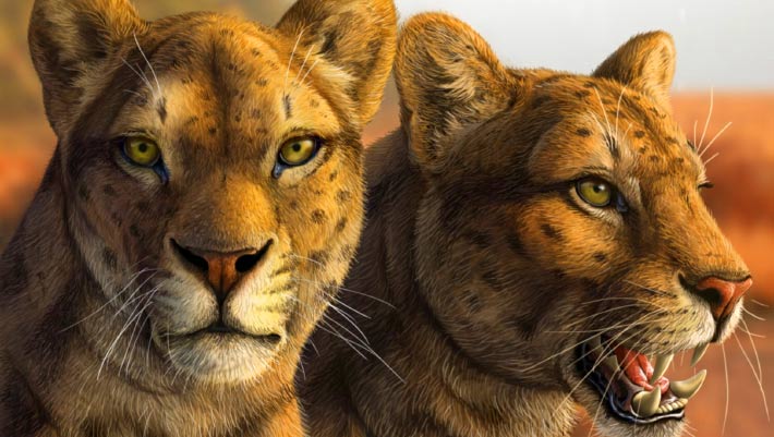
Early Saber-Toothed Cat Gets New Face after Decades of Mystery
Le premier chat à dents de sabre obtient un nouveau visage après des décennies de mystère
dent paléoart reconstitution États-Unis Pliocène mammifères crâne
Un crâne presque complet découvert il y a plusieurs décennies en Arizona a donné aux paléontologues leur aperçu le plus clair à ce jour d'Adelphailurus kansensis, une espèce énigmatique de félidés qui habitait l'Amérique du Nord il y a plus de 5 millions d'années et occupait une des premières branches de l'arbre généalogique des chats à dents de sabre. L'article Le premier chat à dents de sabre obtient un nouveau visage après des décennies de mystère est apparu en premier sur Sci.News : Breaking Science News.
29/06/2026 sci-news ⚙ Traduction automatique
Un superbe crâne de théropode exposé
musée fossile Tyrannosaurus crâne
Lors d'une visite au Museum für Naturkunde (Berlin) nous en avons profité pour photographier le magnifique crâne de Tristan le Tyrannosaurus rex.  Le crâne est l’un des crânes de tyrannosaure les plus complets et les mieux conservés connus de la science.  Les fossiles de T. rex, exposés à côté du crâne, ont été nommés Tristan Otto.  À
12/06/2026 everythingdinosaur ⚙ Traduction automatique
Les scientifiques pensaient les Denisoviens introuvables : cette découverte spectaculaire change désormais tout
os ADN Chine découverte Homo sapiens origine humaine crâne
Pendant quinze ans, les Denisoviens n'ont eu ni visage ni contour. Juste de l'ADN extrait d'un fragment d'os sibérien grand comme un ongle. En 2025, un crâne exceptionnel découvert en Chine a tout changé. Homo longi, surnommé l'Homme dragon, donne enfin un visage à ce groupe humain fantôme. 
06/06/2026 futura-terre
Ce poisson préhistorique pourrait expliquer comment les animaux ont marché pour la première fois sur Terre
Antarctique crâne
Les scientifiques ont examiné l'intérieur du crâne d'un poisson de l'Antarctique vieux de 380 millions d'années, étroitement apparenté aux premiers animaux à avoir marché sur terre, révélant des indices surprenants sur la façon dont la vie a commencé à sortir de l'eau. Grâce à l’imagerie neutronique avancée, les chercheurs ont découvert que Koharalepis jarviki possédait des caractéristiques adaptées pour vivre près de la surface de l’eau, notamment des ouvertures dans son crâne qui auraient pu l’aider à avaler de l’air et un organe sensible à la lumière lié aux rythmes jour-nuit.
25/05/2026 sciencedaily-paleo ⚙ Traduction automatique
1 2 3 4 5 6